Hospital: Hospital Universitario La Paz.
Nº: C2019-567
Aut@r o Autores: A. Diez Tascon, M. Caicoya Boto, F. García, E. Lanz, A.J. Barrios López, M. Martí De Gracia.
Presentación
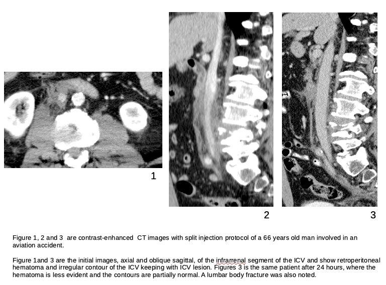
A 66 years old male involved in an aviation accident was brought to our emergency room. The patient presented back pain but preserved the general status. Because of the high energy impact the patient was classified a potentially severe patient and then a full body CT scan with split injection protocol of IV contrast was performed. The findings included retroperitoneal hematoma surrounding the inferior vena cava (IVC) placed under the left renal vein confluence. The contour of this vessel presented abnormalities. A fracture of a lumbar vertebral body was also noted. There was neither evidence of intimal flap nor active extravasation of contrast. The patient was admitted in the critical care unite. The surgical treatment was declined because of the location of the injurie and the preserved status of the patient. After 24 hours a new CT scan was done. The retroperitoneal hematoma had reduced its size and the contours of the IVC had normalized, leading to a spontaneous healing of the injure.
Discusión
The traumatic injuries of the IVC are uncommon but potentially lethal lesions. The mortality rate of these lesions was described to be up to 70%. The most specific findings described in the radiology literature are: Retroperitoneal hematoma, alteration of the contour of the vessel and intimal flap. Other findings described are ICV thrombosis, intracaval fat and hemopericardium. The lethality of the injuries depends on the location. The IVC is divided into four segments with prognostic and therapeutic implications: Supreahepatic, retrohepatic, suprarrenal and infrarrenal. The most severe lesions are those that involve the retrohepatic segment. The distance to the heart is crucial on the prognosis, being the closer lesions to the heart the most severe. The presence of active extravasation of contrast during the CT examination was described also as a worsening prognostic factor.
Conclusión
Inferior vena cava lesions are rare injuries. They should be suspected in the context of acute trauma patients with retroperitoneal hematoma. The severity of this lesion is determined by their distance to the heart and their specific location.
Bibliografía
- Tsai R, Raptis C, Schuerer DJ, Mellnick VM. CT Appearance of Traumatic Inferior Vena Cava Injury. AJR. AMERICAN JOURNAL OF ROENTGENOLOGY, 2016, 207:4:705-711.