Hospital: Ramón y Cajal Universitary Hospital.
Nº: C2019-607
Aut@r o Autores: A. López-Frías López-Jurado, A. Vicente Bártulos, E. García Casado, P. Marazuela García, J.M. Blanc Molina, B. Alba Pérez.
Presentación
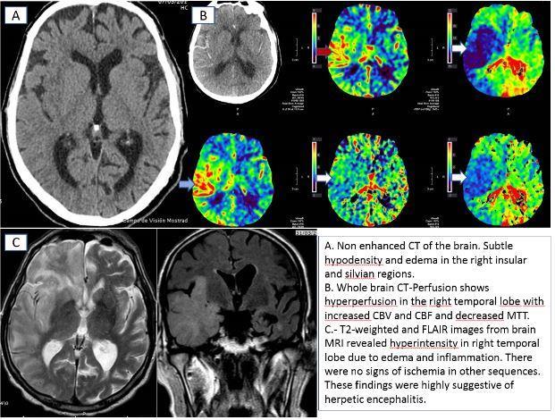
82-year-old-man with a history of liver transplantation and dilated miocardiopathy was admitted our hospital with acute onset of left sided weakness, acute confusion and progressive deterioration of consciousness. A stroke code was activated, and multimodal CT was obtained with the following results: - Basal CT showed subtle right insular hypodensity with mass effect and asymmetry of lateral ventricles. - CT perfusion showed decreased time to peak and mean transit time values, with increase of blood flow and blood volume in the right temporal and frontal lobes. - CT angiogram demonstrated no evidence of vessel occlusion or critical stenosis. Because of deterioration in level of consciousness, the patient was admitted to the ICU department, intubation was inevitable and several treatments including intravenous acyclovir were administered.
Discusión
Emergency department physicians often face the challenging task of differentiating between true acute ischemic stroke and other neurological entities that may show similar neurologic symptoms. These entities are the so-called "stroke mimics", and account for one-fifth from the total of cases when acute stroke is suspected. Imaging may therefore be critical in the acute setting. Unfortunately, non contrast CT is not a sensitive nor specific tool for differentiation, so we must search for other diagnostic tools in order to help us make a more precise diagnosis. There are extensive data supporting the use of CT perfusion in acute stroke management, providing valuable information of brain damage and tissue at risk. Also recent studies have shown its utility characterizing other entities apart from acute stroke. In this case, perfusion CT demonstrated a hyperperfusion area in the right temporal lobe instead of the classic findings of hypovolemia seen in acute stroke. The differential diagnosis included inflammatory-infectious encephalitis, epileptiform activity, and migraine, as well as other more rare pathologies. In our case lumbar puncture was performed to rule out encephalitis an also a MRI study was obtained 2 days after the initial scan. MRI revealed signal hiperintensity in the righttemporal lobe in T2-weighted and FLAIR images, without signs of infarct in the diffusion sequence.
Conclusión
Herpes Simplex Virus Encephalitis with signs of hypervolemia in CT perfusion.
Bibliografía
- Koopman A1, de Leeuw FE2, Meijer F3. CT perfusion hypervolemia: brain ischemia or stroke mimic?. Neuroradiology. 2019 Feb 4. doi: 10.1007/s00234-01902175-3. - Marco de Lucas E1, González Mandly A, Gutiérrez A, Sánchez E, Arná