
Hospital: Hospital Universitario Ramón y Cajal. Servicio de Radiodiagnóstico.
Nº: C2019-148
Aut@r o Autores: L. González-Campo, V. García Blázquez, A. Vicente Bártulos, M. Chiva De Agustín, F. González Tello, J.V. Quintana Pérez.
Presentación
A 54-year-old woman who came to the Emergency Department for pain in the left breast of two weeks of evolution that has been increasing and did not relieve with usual analgesia. In the assessment, the patient was afebrile, the breast was enlarged, painful on palpation, without temperature increase or erythematous appearance and without palpation of axillary adenopathies. The patient reported pain and large increase in breast size in a short time.
Discusión
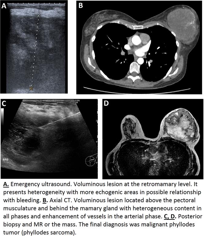
Emergency ultrasound was performed, showing a voluminous, well defined lesion that measured about 7.5 cm in its largest diameter at the retromamary level. It presents heterogeneity with more echogenic areas in possible relationship with bleeding and some areas of vascularity on color Doppler. The possibilities considered were hematoma or abscess, although the existence of some vessels on color Doppler did not allow to rule out a complicated tumor lesion. Finally, it was decided to perform CT to assess the existence or not of bleeding and to characterize the lesion. Basal, arterial and venous scans were carried out. There was a voluminous lesion located above the pectoral musculature and behind the mammary gland with heterogeneous content in all phases and enhancement of vessels in the arterial phase. There was a fatty plane of separation of the pectoral muscles. In view of these findings, the possibility of a complicated tumor lesion was raised and we had to think about the possibility of a sarcomatous lesion with signs of hemorrhage. After further studies (biopsy guided by ultrasound, resonance and pathological anatomy result), the final conclusion of malignant phyllodes tumor (phyllodes sarcoma) was reached. These tumors represent 0.3-1% [1,2] of breast neoplasms. They develop especially in adult women between 35 and 55 years [3] and the typical form of presentation is pain and rapid growth of the breast.
Conclusión
If we find a rapidly growing breast mass we must first assess whether the cause corresponds or not to the breast tissue or the growth has an origin behind the pectoral muscle. Although the first pathology we should think about is the infectious pathology, we must never forget the possibility of tumor pathology and some types of rapidly growing tumors such as malignant phyllodes.
Bibliografía
- Yabuuchi H, Soeda H, Matsuo Y, Okafuji T, Eguchi T, Sakai S, et al. Phyllodes Tumor of the Breast: Correlation between MR Findings and Histologic Grade. Radiology. 2006 Dec 1,241(3):702–9. - Phyllodes tumor
Radiology Reference Article