Hospital: Hospital Clinico San Carlos, Hospital Clínico San Carlos.
Nº: C2019-669
Aut@r o Autores: L. Escudero, L. Galván, H. De La Rosa, L. Pérez.
Presentación
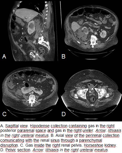
A 66 year old male presented to the ER with a history of 4 day abdominal pain and sweating. On physical exploration he showed intense pain with deep palpation of the right flank, without signs of peritonism. Blood tests revealed elevation of inflammatory parameters, as well as a subtle creatinine elevation. Imaging tests were requested in order to approach this non specific clinical picture. Abdominal ultrasound was performed, detecting a renal fusion abnormality consistent with horseshoe kidney and a right perinephric collection. An optimal exploration was not possible due to the patient’s morbid obesity, the acute pain and the amount of abdominal gas. A contrast-enhanced abdominal CT was performed, acquiring the images in a portal phase. It showed a wide collection containing hipodense fluid and gas in the right perirenal space, extending to the posterior pararenal space and communicating with a disrupted renal parenchyma and likely with the dilated right renal pelvis, also containing gas. An 8 mm lithiasis inside the right ureteral meatus was detected. Difuse urothelial enhancement, left retrocrural lymphadenopaties and a small amount of free fluid in the right paracolic space were also present. The findings were consistent with emphysematous pyelonephritis. The lithiasis was visualized and extracted endoscopically, and a JJ catheter was placed in the right urether. After two weeks of torpid clinical evolution, percutaneous TC guided drainage of the perinephric abscess was performed, but two-months later, an open right nephrectomy was necessary because of clinical deterioration and urin leak persistence on CT.
Discusión
Emphysematous pyelonephritis is a severe necrotizing affection of the renal parenchyma and surrounding tissues, strongly associated with diabetes, which can be caused by obstructive uropathy. Clinical and laboratory features are not likely to lead to an early diagnosis, therefore, imaging becomes essential. Since ultrasonographic exploration can be challenging, doubtful cases should get a complementary CT. The most commonly involved pathogens are E. Coli (most cases), Klebsiella, Proteus and Pseudomona. Deterioration of general condition and poor response to convencional antibiotics demand an early management. Horseshoe kidney is the most common congenital renal fusion anomaly and is usually an accidental finding. When symptomatic, hydronephrosis, infection or lithiasis are common causes.
Conclusión
A horseshoe kidney is not likely to present with clinical manifestations, but when it does, a picture of lithiasic uropathy or infection is expected. Emphysematous pyelonephritis is difficult to suspect clinically, therefore, imaging becomes fundamental in its diagnosis and management. This severe condition may lead to a torpid clinical evolution with poor therapeutic results.
Bibliografía
- A. BLANCO DÍEZ, A. BARBAGELATA LÓPEZ, E. FERNÁNDEZ ROSADO, R. CASAS MUÍÑO, V. CHANTADA ABAL, M. GONZÁLEZ MARTÍN. Pielonefritis enfisematosa: presentación de un caso y revisión de la literatu