Hospital: Complejo Asistencial Universitario de Palencia.
Nº: C2019-670
Aut@r o Autores: J. Cuello Ferrero, M. Ruiz Martín, K. Lakis Granell, E. De Sande Nacarino, J. Simal Fernández, B. Viñuela Rueda.
Presentación
A woman in her sixties came to the ER with a fever, severe cervical pain, disphagia and disphonia. Acute bacterial thyroiditis was suspected.
Discusión
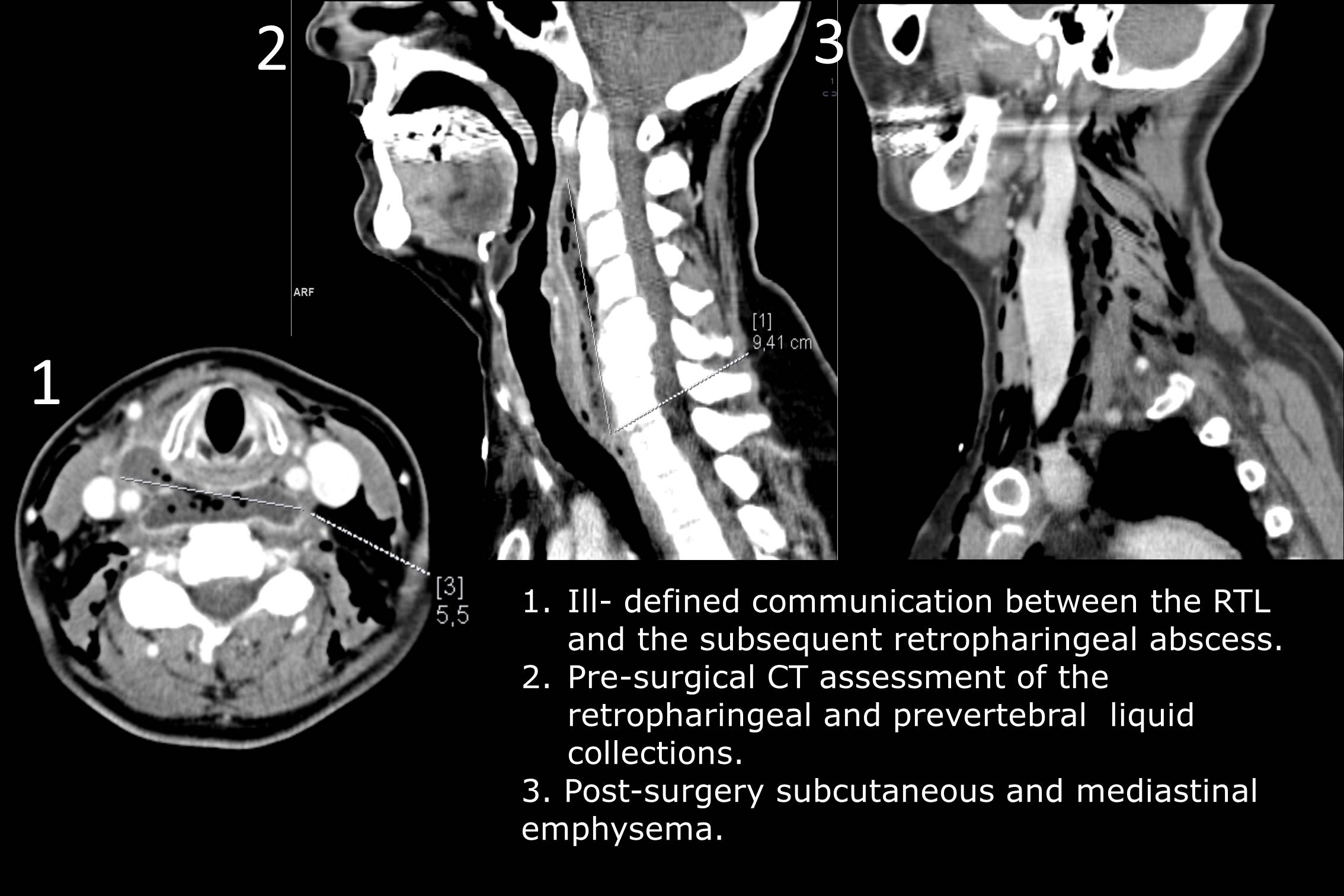
An US examination of the gland was carried out. Using this technique a nonhomogenous thyroid with some pseudo-nodular hipoecogenic patches affecting the right thyroid lobule (RTL) was identified. On the suspicion that an infectious thyroiditis was taking place, an enhanced-CT of the neck followed. A non-homogenous enhancing nodule on the RTL linked to a retropharingeal liquid collection was reported. Its extension ranged from the margin of the RLT to the anterior aspect of the carotid space and the right lateral visceral space, both of them full of liquid content with gas bubbles within. Some laterocervical reactive lymph nodes adjacent to the capsulated collection were present. There were no other significant lesions. The findings were highly suggestive of acute suppurative thyroiditis with secondary retropharingeal and prevertebral abscesses. After antibiotic treatment was set, surgical drainage by cervicotomy was carried out by head-neck-and-ear surgeons, placing a drainage tube to ensure removal of the collected fluid. A follow-up CT revealed moderate subcutaneous and mediastinal emphysema after the procedure. A second surgical procedure was performed to broaden the area of drainage and place a second drainage tube so the purulent content would be vacated more easily. The postoperative CT showed a small number of remaining intraparenchimal infectious foci while mild inflammatory changes took over the affected area, pathology reports of the fluid showed no evidence of bacterial growth. A full antibiotic course was applied until resolution of symptoms. On the basis of these findings a diagnosis of auto-immune thyroiditis was proposed. Follow-up visits were planned until full recovery.
Conclusión
Knowledge of the visceral spaces of the neck is vital for proper assessment of infectious/suppurative diseases of the head, neck and its contents (thyroid, sallivary glands, lymph nodes...). Follow-up CT are necessary to assess the progression of visceral infections such as suppurative thyroiditis. Given the anatomical complexity of the area US or CT are useful tools in suspecting and diagnosing infectious pathology of the head and neck area.
Bibliografía
- Gonzalez-Beicos A, Nunez D. Imaging of Acute Head and Neck Infections. Radiologic Clinics of North America [Internet]. 2012 [cited 2 April 2019],50(1):73-83.Available from: https://www.clinicalkey.es/#!/content/journal/1-s2.0S0033838911001527 - Harnsb