Hospital: HCUVA
Nº: C2019-317
Aut@r o Autores: G. De Paco Tudela, M. Ato Gonzalez, A. Navarro Baño, P. Rey Segovia, D. Gea Martos, A. Castillo García.
Presentación
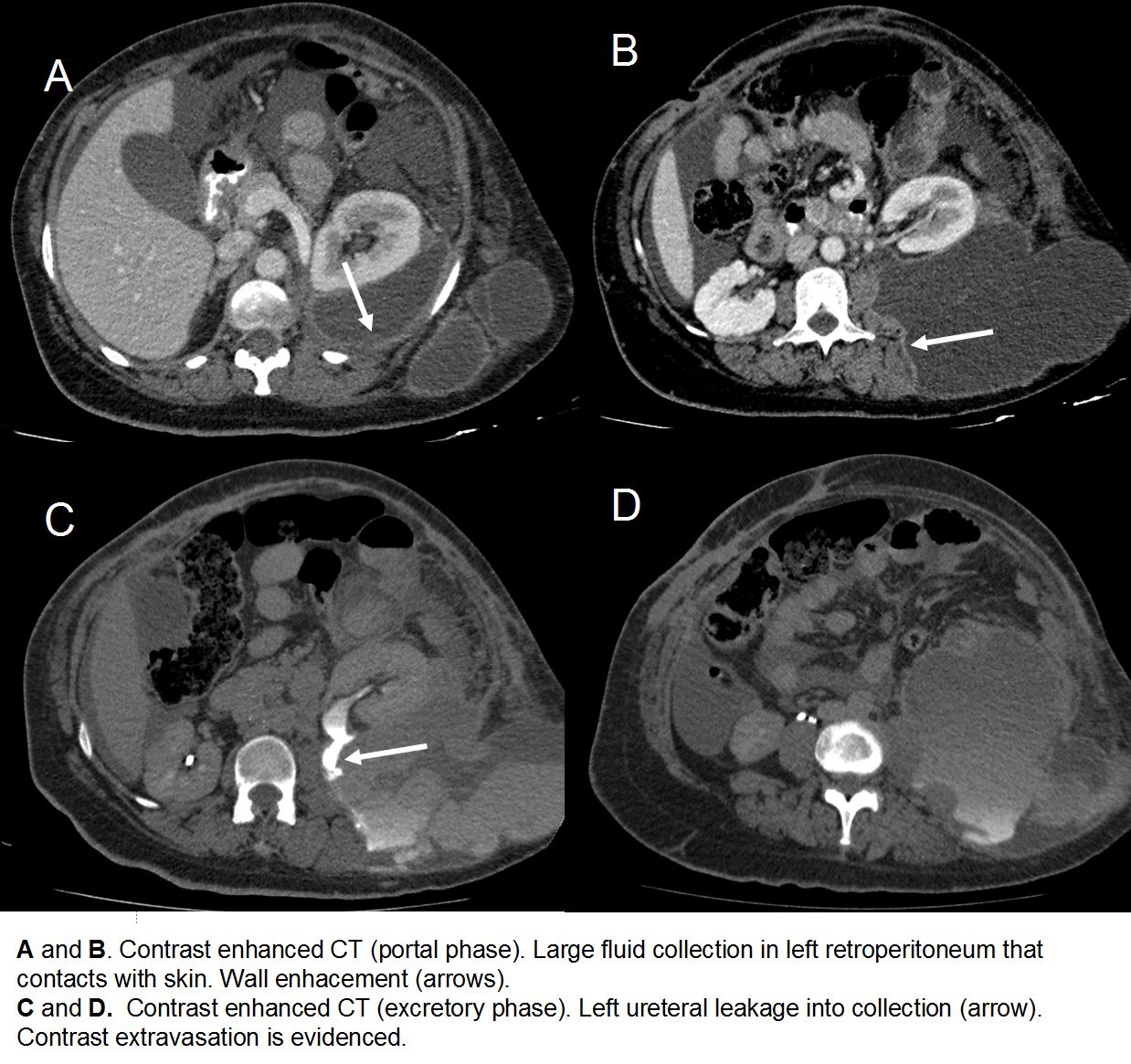
A 55 year old women with a history of hyperthension who presented with diffuse abdominal pain. She had a pheochromocytoma removal surgery 1 month before, without inmediate complications. At admission, patient showed diffuse abdominal pain, especially in hypogastrium and left flank, refractory to habitual analgesia, and absence of bowel movements for 4 days, without nausea or vomits. The patient arrived tachycardic with leukocytosis. A contrast-enhanced two phase abdominal CT with portal and excretory phases was performed, showing a left ureter leakage (Figure 1.C , arrow) into a large fluid collection in left retroperitoneum that pushes anteriorly the kidney and extends to the subcutaneous cellular tissue contacting with the skin. It shows wall contrast enhacement (Figure 1.B, arrow). This findings are suggestive of infected urinoma. The following day the collection was drained and a nephrostomy was performed. The following week it was surgically repaired.
Discusión
Urinoma is a rare pathology that consist of urine colections that are usually found in the retroperitoneum, oftenly in the perirenal space. The urine leakage can be secondary to obstruction of the urinary tract, trauma or iatrogenic post-surgery lesion of the urinary tract. The clinical presentation is inespecific, e.g abdominal pain, hematuria, or fever in case it's infected. If the patient has been surgically operated, especially a urologic intervention, the diagnostic of yatrogenic urinoma must be taken into consideration. A contrast enhanced CT must be performed, always including an excretory phase in order to discover urinary extravasation. Small perirenal urinomas can be treated conservatively, but in case of large size, hydronefrosis, fever, compression or demonstration of urinary tract lesion, prompt surgical repair must be performed in order to prevent complications as abscess, hydronephrosis or loss of renal function.
Conclusión
Urinoma must be suspected when a patient with trauma or recent surgery presents abdominal pain. CT must be performed, always including an excretory or delayed phase in order to demonstrate urinary extravasation.
Bibliografía
- Yang DM, Jung DH, Kim H, Kang JH, Kim SH, Kim JH, Hwang HY: Retroperitoneal cystic masses: CT, clinical, and pathologic findings and literature review. Radiographics. 2003, 24: 1353-1365. - Jou YC, Shen CH, Cheng MC, Lin CT, Chen PC. Bilateral ureteral