Hospital: Hospital Ramón y Cajal.
Nº: C2019-617
Aut@r o Autores: R. Romera Sánchez, I. García Gómez Muriel, C. Sempere Ortega, J. Blanc Molina, L. González Campo, C. Picón Serrano.
Presentación
A 63-year-old male with no history of interest, who comes for discomfort in both flanks and epigastric pain, with no other symptoms. Laboratory parameters show elevation of C-reactive protein, LDH with slight increase of bilirubin and transaminases. If cholangitis is suspected and the pain does not improve clinically, an imaging test is requested.
Discusión
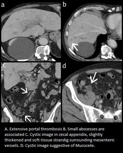
We first performed an ultrasound, the only finding was an extensive portal thrombosis, the bile duct was not dilated, and no other findings were found. We decided to complete the study with CT with intravenous contrast confirming the thrombosis affecting the main portal and both branches. Thrombosis extended through the upper mesenteric vein and small mesenteric vessels of right iliac fossa accompanied by fat stranding.In addition, in the hepatic parenchyma there was a small nodular lesion with poorly defined edges. These findings made it necessary to rule out pylephlebitis with small hepatic abscess, or tumour thrombosis with hepatic metastasis. We opted for the first option as most likely already in slight thickening of the appendix walls, with an associated cystic image, data that could be related to superinfected mucinous neoplasia. Diagnosis that was confirmed in surgery once the acute clinical event was resolved. Portal thrombophlebitis, pylephlebitis, is usually secondary to infections in regions drained by portal venous system. The most frequent cause is diverticulitis, but other pathologies are also included such as appendicitis, urinary pathology, pelvic pathology, pancreatitis... The clinical manifestations may be asymptomatic, exhibit minimal symptoms related to the primary infection site, or have an acute abdomen. The most frequent initial manifestation of appendiceal tumors is acute appendicitis. Epithelial neoplasms are the most frequent along with neuroendocrine tumors. The classic radiologic manifestation of an appendiceal mucinous tumor is a mucocele, macroscopic description of a mucin-dilated appendix. At CT, a mucocele manifests as a dilated appendix filled with homogeneous low-attenuation material.
Conclusión
It is important to think of non-tumour pathology or infectious complications in the case of portal thrombosis as this changes the management of the patient. As for appendicular tumors that manifest as appendicitis, it is also important to consider their differential diagnosis since the surgical approach may be different.
Bibliografía
- García R, Liñares M, Baleato S, Villalba C. Pylephlebitis Radiology: Volume 255: Number 3—June 2010 - .Duran R, Denys A, Letovanec I, Meuli R, Schmidt S. Multidetector CT Features of Mesenteric Vein Thrombosis RadioGraphics 2012, 32:1503–