Hospital: Hospital Universitario Virgen de las Nieves.
Nº: C2019-274
Aut@r o Autores: J. Parejo, A. Martinez.
Presentación
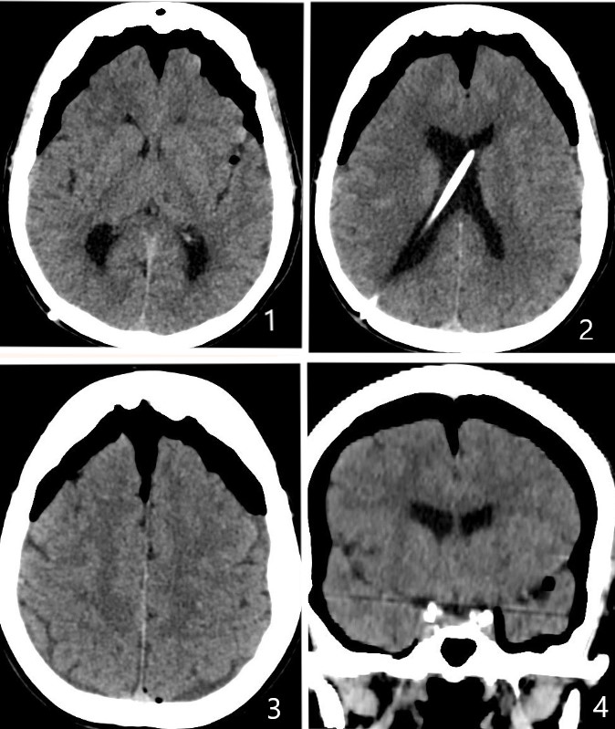
A 76 years old women was taken to the emergency department with progressive headache, bradypsychia, decreased alertness and general weakness. She had been hospitalised 5 days ago for intracranial pressure (ICP) monitoring due to suspicion of ventriculoperitoneal (VP) shunt malfunction. This patient had a VP shunt since 10 years ago for treatment of normal pressure hydrocephalus. Her past medical history was also significant for hypertension and toxic nodular goiter.She was request for an emergency head TC. It showed plentiful amount of trapped intracranial gas resulting in mass effect, an entity known as tension pneumocephalus.
Discusión
As a result of this, we have reviewed in the literature the cases of tension pneumocephalus as a complication of ICP monitoring, as well as the CT radiological findings that allow us to suspect this entity. As far as we know, only two cases of this entity have been associated with PIC monitoring (one of them with traumatic brain injury and the other with non-traumatized patients).Intracranial pressure monitoring (ICP) is a relatively common procedure. It can trigger complications such as infection or hemorrhage. However, the tension pneumocephalus is infrequent and needs to be taken into account given the seriousness involved. The term pneumocephalus defines intracranial air. It occurs when air that enters through a dural defect is unable to escape. When it occupies a large volume can exert compression on the brain tissue and cause brain herniation, so it is considered a neurosurgical emergency that requires decompression. In TC it is distinguished as hypodense collections of very low attenuation (around - 1000 HU) in the subdural space (images 1-4). Care needs to be taken in ensuring that it is not fat which although of much higher density (-90HU) also appear completely black on routine brain windows. Initially, the air externally compresses both frontal lobes toward the midline (peak sign). Finally if the air separates both frontal lobes it gives a typical image (sign of Mount Fuji), as seen in images 1-3 .
Conclusión
#¿NOMBRE?
Bibliografía
- Heaney JA, Peter Gan YC. Tension Pneumocephalus as a Complication of Intracranial Pressure Monitoring. Ann Clin Case Rep. 2017, 2: 1266. - Pulickal GG, Sitoh YY, Ng WH. Tension pneumocephalus. Singapore Med J. 2014,55(3):e46–e48.