Hospital: Hospital de 12 Octubre.
Nº: C2019-600
Aut@r o Autores: I. Ríos Gómez, I. Navas, L. Ibañez Sanz.
Presentación
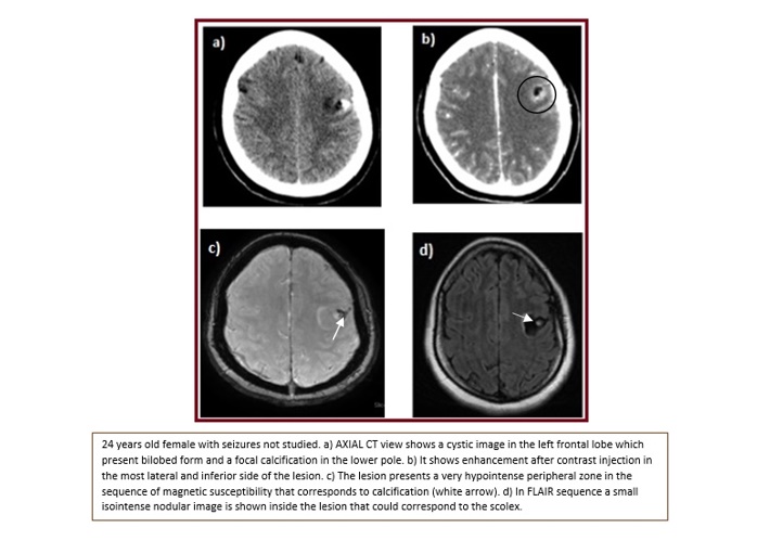
24 years old female with seizures not studied. CT examination showed a cystic lesion on the left frontal lobe, which presents bilobed shape and a focal calcification in the lower pole. It causes a certain mass effect on the adjacent convexity suci. It shows enhancement after contrast injection in the most lateral and inferior side of the lesion. On MRI, the lesion presents a very hypointense peripheral zone in the sequence of magnetic susceptibility that corresponds to calcification on CT. The CIV administration shows the peripheral enhancement in the lateral and inferior margin. In the T2 flair sequence, a small isointense nodular image is shown inside the lesion that could correspond to the scolex.?
Discusión
Cysticercosis is a Taenia solium infection. When it involves the CNS, it is called neurocysticercosis (NCC). Extra-intestinal disease occurs as a result fecal-oral contamination. Neurocysticercosis is the most common parasitic infection of the brain and the mean cause of epilepsy in the developing world, especially Latin America. Clinical presentation includes: seizures, headaches, altered mental status, neurological deficits… There are four main stages of presentation: vesicular, colloid vesicular, granular nodular and nodular calcified. The first is the vesicular a cyst with a translucent vesicular Wall and transparent fluid. In the colloidal stage, the vesicular cyst develops a thick wall, the fluid becomes turbid. As in our case, the cyst continues to degenerate as it moves into the granular stage which is characterized by a thick wall, degenerated scolex and inflammatory process decrease. At this stage, they show enhancement after contrast injection. In T2 and FLAIR sequences there is perilesional hyperintensity in relation to vasogenic edema. Diffusion sequences show variable intensity and hyperintensity in ADC. Lastly, increasing evidence implicates calcified NCC in the development and maintenance of seizures and epilepsy. In population-based studies calcified lesions on CT are much more common than viable cysts, and they are more prevalent in patients with epilepsy than they are in asymptomatic patients.?
Conclusión
Neurocysticercosis is the most common parasitic infection of the brain and the most common cause of seizures in young adults in endemic areas. CT has a high sensitivity and specificity in most forms of neurocysticercosis and is superior to MR imaging in identifying calcifications. However, the main advantage of MR imaging over CT is its higher contrast resolution, particularly helpful in the evaluation of ventricular involvement and the detection of inflammatory changes.
Bibliografía
- De Giorgio CM, Medina MT, Duron R, Zee C, Escueta SP. Neurocysticercosis. Epilepsy Curr. 2004, 4:107–1 - Costantino SA, Capiel CA, Rossini SA, Landi M, Bouzas CA. Diagnostic imaging in neurocysticercosis. Rev Arg Diag Por Imágenes. 2012, 1:24– 3