Hospital: Hospital Universitario Alava.
Nº: C2019-334
Aut@r o Autores: L. Alonso Irigaray, A. Sanchez Garcia, I. Hernandez Sastre, A. Valero Macia, P. Rosado Caracena, E. Diez Lasheras.
Presentación
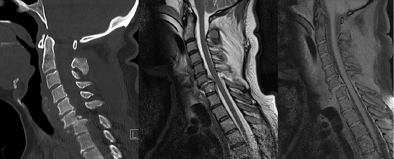
45 year old female, 36 weeks pregnant (multifetal pregnancy), who is transferred to our center after a high speed traffic accident. At her arrival she is stable, Glasgow 15. Refers cervical and dorsal pain, headache and abdominal pain at left hypochondrium. An abdominal ultrasound is performed at the emergency box that shows integrity of the placenta and normal fetal heart rate. There is no evidence of damage in abdominal organs and no ascites. As the headache persist, a non contrast CT is performed including head, neck and thorax: . - Brain: no significant findings. . - Neck: bilateral interfacetal fracture-dislocation of C6-C7 with grade 1-2 anterolisthesis and indirect signs of instability / fracture of posterior ligament complex. C7 anterior - superior corner fracture with anterior displacement of the fracture fragment ans C6 posterior - inferior corner with posterior displacement of the fragment. . - Thorax: fracture of the sternum body, bilateral pleural effusion, small pulmonary contusions at upper right hemithorax. In consensus with traumatologists and gynecologists, the patient underwent an urgent caesarean and right after that, a cervical an dorsal MRI to evaluate the integrity of the spinal cord. MRI confirms CT findings: bilateral interfacetal fracture-dislocation of C6-C7, fracture of posterior ligament complex on C6-C7, fracture of intervertebral disc on C6-C7, no spinal cord damage and absence of epidural hematoma. An spinal surgery is performed: closed reduction and primary arthrodesis under general aesthesia. There weren´t any complications during the procedure and the post-surgical evolution was satisfactory.
Discusión
Cervical spine injuries occur in 5%–10% of patients with blunt polytrauma and are classified under the harmful mechanism, but the estability of the lesion establishes its severity and therefore the need for surgical treatment. The SLIC classification is used to evaluate the risk of spinal cord damage in cervical fractures from C3 to C7 and is based on 3 components of injury: morphology of the lesion, integrity of the posterior ligamentous complex and neurological status of the patient. In our case, the damage is caused by a hyperflexion mechanism, causing bilateral interfacet dislocation, fracture os posterior ligament and no spinal cord injury.
Conclusión
CT is used as the initial screening examination for patients who are suspected of having cervical spine trauma and is often enough for making the determination of whether surgery is necessary or not. There are others determinants of management, including disk herniations, ligamentum flavum infolding, cord damage and epidural hematoma, are not well evaluated with CT but are clearly depicted at MR imaging.
Bibliografía
- Multidetector CT of Blunt Cervical Spine Trauma in Adults. RadioGraphics 2014, 34:1842–1865. Dreizin, D. MD and cols. - Imaging of Adult Cervical Spine Trauma. RadioGraphics 1988, Voulme 8, Number 4, Pages 667-694. Berquist, T.H.