Hospital: Hospital Universitario Ramón y Cajal.
Nº: C2019-681
Aut@r o Autores: V. García Blázquez, A. Vicente Bártulos, J. Pérez-Templado, M. Andreu Rodríguez, A. Silva, M. Chiva De Agustín.
Presentación
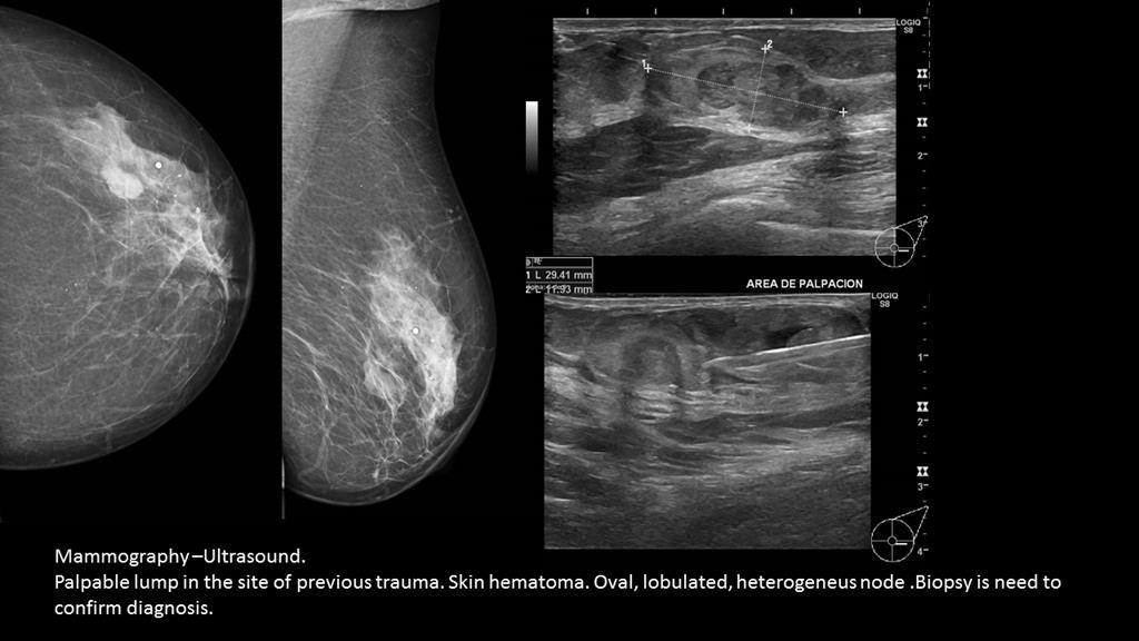
A 74 years old woman, with any previous pathology, was admitted to the emergency room (ER) for finding, after a self-examination, a possible node in left breast, performed after a costal trauma with a skin hematoma in the breast. The patient referred pain mild bilateral costal bones. An ultrasound was performanced with initial diagnosis of hematoma and she was refered to Breast Radiology Unit at our Hospital one week after the episode. Mammography and a ultrasoud showed a partially defined heterogeneous node of 30 mm, oval, lobulated, without microcalcifications in the lower outer quadrant of the left breast which, associated with the clinical features, seemed to be an hematoma. It was recommended a new ultrasoud one month later. Because of the stability of the node features, a biopsy was taken out with result of malignity: “invasive mucinous carcinoma”.
Discusión
The mammary gland is a common site for traumas which often lead to the formation of intraglandular hematomas. Clinically, these patients may be asymptomatic or may present with a palpable lump, skin tethering, induration, and occasionally axillary lymphadenopathy. Depending on the time at which diagnostic imaging is performed, hematoma can have highly variable appearances. Mostly, are easily diagnosed by both mammography and US. However, the diagnostic features are difficult to distinguish from malignant breast nodules. Self-examination after a causative trauma, incidentally can discover malignat nodules. Also, mucinous carcinoma of the breast is one of the rarer forms of intramammary cancer, often presenting as a lobulated, fairly well circumscribed mass on mammography and sonography, which can mimic benign lesions.
Conclusión
Breast node must always be thought of as a differential diagnosis for a breast hematoma, regardless of previous trauma. A specialist in breast radiologist must be who valued the lesions in the breast and who determine the need for biopsy.
Bibliografía
- Tayyab SJ, Adrada BE, Rauch GM, Yang WT. A pictorial review: multimodality imaging of benign and suspicious features of fat necrosis in the breast. Br J Radiol. 2018 Dec,91(1092):20180213. doi: 10.1259/bjr.20180213. Epub 2018 Jul 31. - Kelli Y. Ha, MD,