Hospital: Hospital Universitario de Puerto Real.
Nº: C2019-375
Aut@r o Autores: A. Luna Morales, M. Sanchez - Porro Del Río, M. Pérez Benítez, E. García Gámez.
Presentación
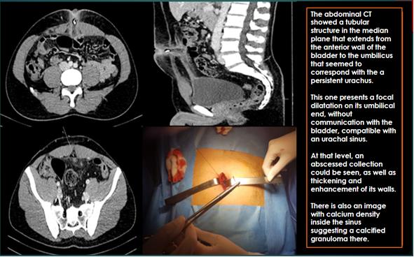
A 34-year-old man, who suffered fever, umbilical pain and omphalitis, with poor evolution despite antibiotic treatment. He presented an erythematous lesion in the umbilical region with significant perilesional cellulitis and foul-smelling purulent discharge from the umbilicus. An abdominal CT was requested to evaluate the need of an urgent treatment. The abdominal CT showed a tubular structure in the median plane that extends from the anterior wall of the bladder to the umbilicus that seemed to correspond with the a persistent urachus. This one presents a focal dilatation on its umbilical end, without communication with the bladder, compatible with an urachal sinus. At that level, an abscessed collection could be seen, as well as thickening and enhancement of its walls. There is also an image with calcium density inside the sinus suggesting a calcified granuloma there. The Surgeons performed a dissection and ligature of the urachal remnant.
Discusión
The urachal sinus corresponds to one of the less frequent urachal anomalies (15%), originated when the umbilical end of the urachus does not close and persists as a fusiform structure just below the umbilicus. This anomaly of the urachus represents a potential space in which the accumulation of cellular debris favors the development of complications, such as infection and lithiasis formation. The US, CT and MR images reveal a thickened and fusiform blind dilatation of the urachus at the umbilical end without communication with the bladder. The majority of patients with urachal pathology, (except persistent urachus), are asymptomatic. However, they become symptomatic if they develop an infection and other complications. The approach to patients with urachal anomalies has evolved over time and remains controversial. Although symptomatic patients have traditionally been treated surgically, nowadays some authors use a new algorithm that proposes a treatment depending on some facts like the age or the symptomatology of the patient.
Conclusión
Abnormalities of urachus in adults can cause considerable morbidity and potential mortality. The clinical diagnosis is often delayed due to nonspecific symptoms or lack of them. Usually, the US is used for an initial evaluation, and CT or MR images are used to further characterize and evaluate the possible complications, mainly infections and the development of malignant tumors, as well as to help optimize an adequate surgical approach if necessary.
Bibliografía
- Parada Villavicencio C, Adam SZ, Nikolaidis P, Yaghmai V, Miller FH. Imaging of the Urachus: Anomalies, Complications, and Mimics. Radiographics: a review publication of the Radiological Society of North America, Inc. 2016 Nov, 36 (7): 2049-2063. - Yu