Hospital: Hospital Universitario Virgen de la Arrixaca.
Nº: C2019-131
Aut@r o Autores: P. Rey Segovia, Á. Cepero Calvete, A. López Sánchez, M. Abellan Rivero, D. Gea Martos, G. De Paco Tudela.
Presentación
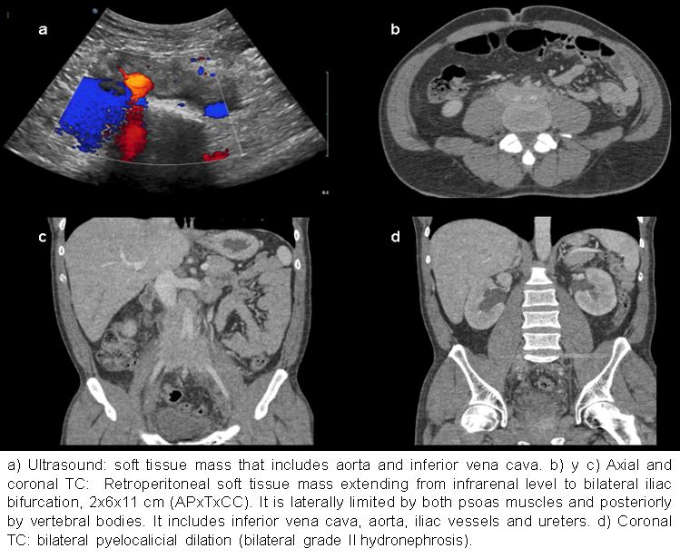
A 37-year-old man was attended at emergency department due to episodes of right lumbar pain irradiated to mesogastrium, accompanied by nausea and polyuria. In the blood analysis high creatinine (1.85) and PCR levels were detected. Abdominal ultrasound was performed demonstrating the existence of bilateral grade II hydronephrosis, as well as a soft tissue mass that included the inferior vena cava, aorta and ureters. Regarding to the previous findings, an abdominal CT with intravenous contrast was done. The CT confirmed the existence of a retroperitoneal mass, measuring 2x6x11 cm (APxTxCC), extending from the infrarrenal level to the iliac bifurcation, enveloping the ureters, aorta and inferior vena cava, without significant displacement of the referred structures. Lymphoma was presented as the first diagnostic option without being able to rule out other etiologies such as retroperitoneal fibrosis or Erdheim-Chester disease. The biopsy performed reported that the morphological findings and immunophenotypic results could be correlated with retroperitoneal fibrosis.
Discusión
Retroperitoneal fibrosis (RPF) is a rare collagen vascular disorder of unclear cause. It is typically seen in people 40–60 years old with more prevalence in men. Most cases are thought to be idiopathic, the remainder occur in association with inflammatory disorders, malignancies, or medications. The initial signs and symptoms are often nonspecific, such as malaise, anorexia, low-grade fever, and poorly localized pain over the flank. As the degree of fibrosis progresses, the symptoms are mainly related to entrapment and compression of retroperitoneal structures, frequently entrapping and obstructing the ureters. Ureteral involvement is bilateral in most cases. Multidetector CT and MR imaging, has become the mainstay of noninvasive diagnosis of RPF. It allows comprehensive evaluation of the morphology, location, and extent of RPF and involvement of adjacent organs and vascular structures. The typical morphologic findings consist of a well-delimited but irregular soft-tissue periaortic mass, which extends from the level of the renal arteries to the iliac vessels and often progresses through the retroperitoneum to envelop theureters and inferior vena cava. The mass usually lies anterior and lateral to the aorta, sparing the posterior aspect and not causing aortic displacement.
Conclusión
The most important diagnostic challenge is differentiation of benign from malignant RPF. Imaging plays a key role in diagnosis of RPF. It allows evaluation of the location, extent of the fibrosis and complications of this disease process.
Bibliografía
- Cronin CG, Lohan DG, Blake MA, Roche C, McCarthy P, Murphy JM. Retroperitoneal fibrosis: a review of clinical features and imaging findings. AJR Am J Roentgenol. 2008,191 (2): 423-31. - Caiafa RO, Vinuesa AS, Izquierdo RS, Brufau BP, Ayuso Colella JR,