Hospital: Hospital Virgen de la Arrixaca.
Nº: C2019-126
Aut@r o Autores: A. Cuélliga-González, G. Litrán-López, F. Barqueros-Escuer, J. Felices-Farias, D. Páez-Granda, G. De Paco-Tudela.
Presentación
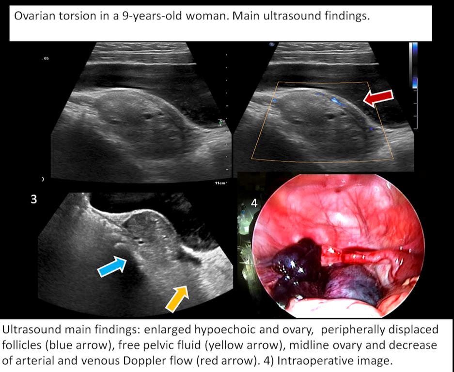
A 9-year-old woman came to our service with abdominal pain. Her mother refferred that the instauration of the symptomatology established five days ago, and since then, the pain progressively increased. There was no nausea, vomiting or fever. Because of the clinical suspicion of acute abdomen, a pelvic-abdominal ultrasound was performed. In this study a normal cecal appendix was identified. Nevertheless some inflammatory changes, as a scarce laminae of free pelvic fluid was observed in the right iliac fossa. This finding was accompanied by an enlarged midline ovary with mixed signal intensity, peripherally displaced follicles, and little intra-ovarian Doppler flow. These findings were reported as an ovarian torsion, that was confirmed after surgery.
Discusión
Ovarian torsion refers to the rotation of the adnexal and portion of the fallopian tube on the supplying vascular pedicle, and is the fifth most common gynecologic surgical emergency. Most patients present with severe non-specific lower abdominal and pelvic pain. This pathology predominates in young women (15-30 years old, 20% during pregnancy) and post-menopausal women, and the principal cause is the presence of an adnexal mass. Torsion of a normal ovary more commonly occurs in young children, as in our case, when developmental abnormalities predispose to his twist. Ultrasound is the initial imaging modality of choice, and the main findings are: enlarged hypoechoic and ovary (>5cm), peripherally displaced follicles, free pelvic fluid: (>80% of cases), midline ovary and decrease of arterial and venous Doppler flow. All of these findings were present in our case. It is very important ti perform an early detection of adenexal torsion, because patients should promptly treated by surgery, to prevent ovarian necrosis.
Conclusión
Ovarian torsion is the fifth most common gynecologic surgical emergency. Ultrasound is the modality of choice, and its main findings must be known by the radiologist, since we could help to prevent an ovarian necrosis.
Bibliografía
- Chiou SY, Lev-toaff AS, Masuda E, Feld RI, Bergin D. Adnexal torsion: new clinical and imaging observations by sonography, computed tomography, and magnetic resonance imaging. J Ultrasound Med. 2007,26 (10): 1289-301. - Kokoska ER, Keller MS, Weber TR.