Hospital: H U N S C.
Nº: C2019-27
Aut@r o Autores: M. Fdez. Del Castillo Ascanio, C.A. Marichal Hernández, V. Martín García, R.D. Medina Herrera.
Presentación
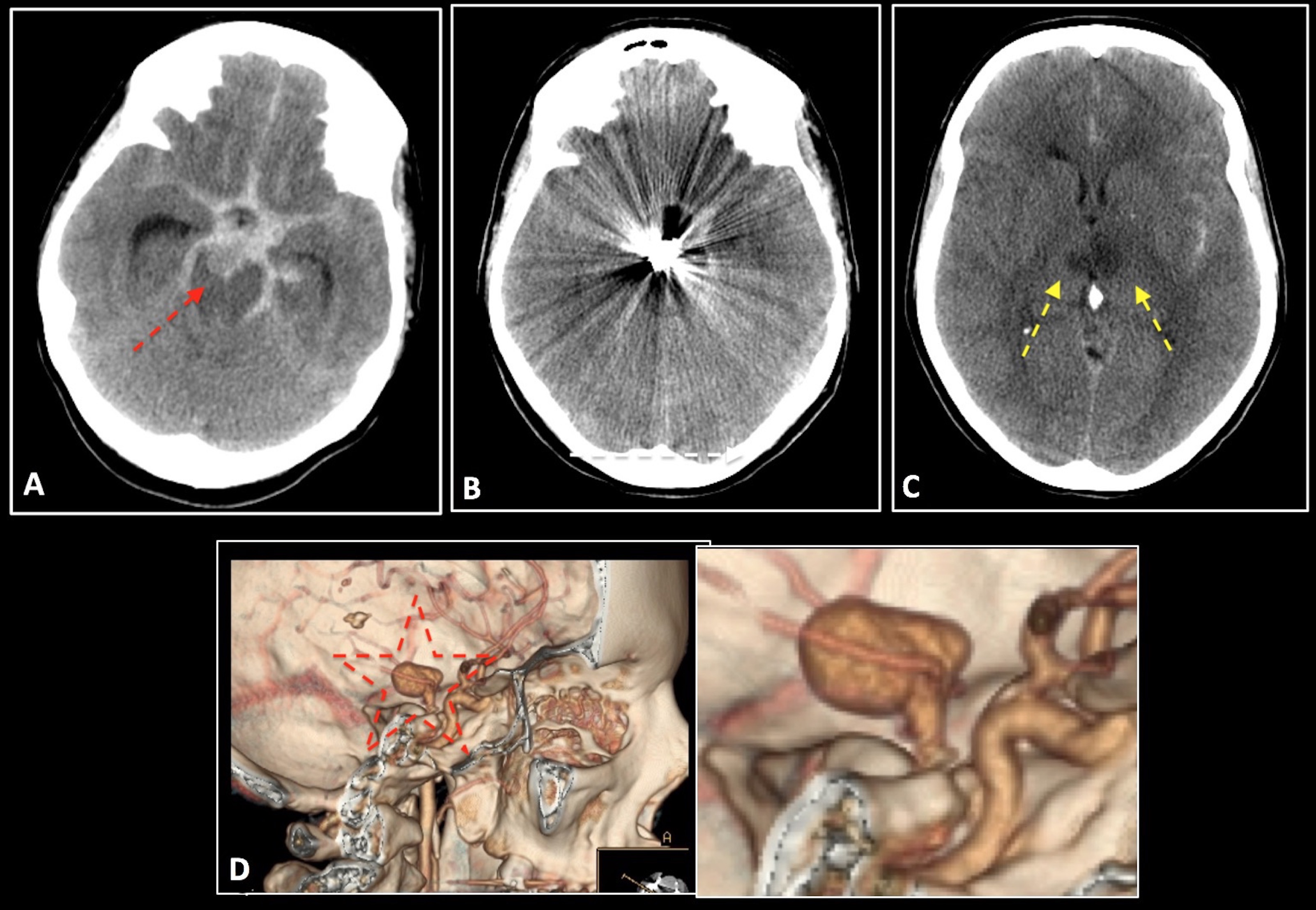
A 54-year-old woman with an intense headache came to the hospital. On suspicion of subarachnoid haemorrhage (SAH), a computer tomography (CT) scan is performed confirming the clinical suspicion of SAH (modified scale Fisher 4). CT angiography is performed where an aneurysm at the top of the basilar is found, and it´s embolized. A few days later, a control CT scan was requested in which bilateral thalamic hypodensity was obtained in the context of ischemic lesions, because of the involvement of the Percheron´s artery.
Discusión
Basilar artery aneurysms are less common than anterior circulation aneurysms and rupture less frequently, but their critical location necessitates careful evaluation. Typically, the haemorrhage of a rupture basilar aneurysms is localised in the interpeduncular cistern but may extend into the suprasellar cistern as in our case. This kind of aneurysms can be treated via endovascular, but have higher rates of progression and retreated progression than other aneurysm locations, independent of treatment modality. Rupture status is associated with increased rates of progression, retreatment, and retreated progression, and endovascular intervention is associated with shorter time intervals to this events. Our patient evolved badly because she probably had an artery of Percheron. The thalamus is supplied predominantly by multiple small branches arising from the ipsilateral posterior communicating artery and P1 and P2 segments of the posterior cerebral artery. Artery of Percheron is an uncommon anatomic variant, in which a single, unpaired thalamo-perforating artery trunk arises from the first part of the posterior cerebral artery and supplies bilateral medial thalami. Occlusion of this artery causes a characteristic pattern of ischemia involving paramedian thalami2. Probably, the proximity of the artery of Percheron origin to the embolized aneurysm led to the ischemic event.
Conclusión
It is important to know the anatomical variants of the polygon of Willis in order to act correctly from a diagnostic and therapeutic way. CAPTION A. Axial CT showing thick subarachnoid haemorrhage with image suggestive an aneurysm at the top of the basilar artery (red arrow). B. Metallic artifact due to embolization material. C. Thalamic bilateral hypodensity suggesting ischemic Percheron´s artery (yellow arrows). D. Vascular reconstruction showing top basilar aneurysm (red star).
Bibliografía
-Sekhar LN, Tariq F, Morton RP, Ghodke B, Hallam DK, Barber et al. Basilar Tip Aneurysms: A Microsurgical and Endovascular Contemporary Series of 100 Patients. Neurosurgery. 2013 Feb,72(2):284-98, discussion 298-9. doi:10.1227/NEU.0b013e318279795 - Abeca