Hospital: Hospital Universitario Bellvitge.
Nº: C2019-504
Aut@r o Autores: K. Pérez Alfonso, M. Serra Salas, A. Güell Bara, D. Nova Vaca, M. Pérez Rubiralta, O. Gasulla Montardit.
Presentación
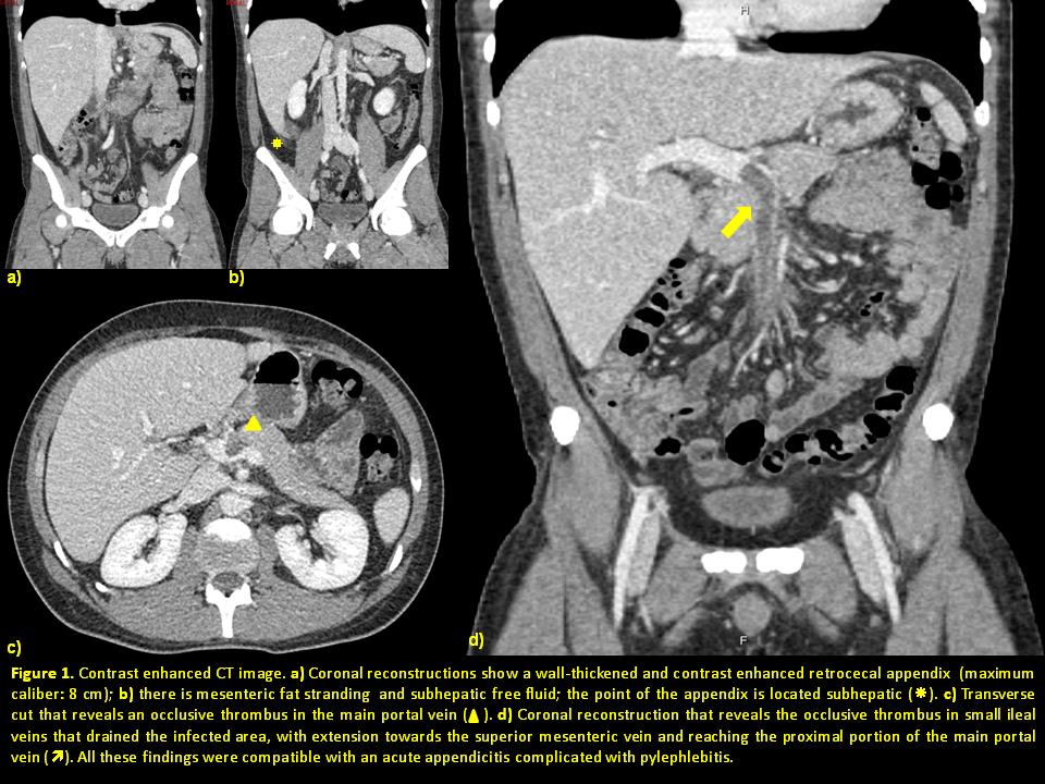
A 39-year-old male presented to the emergency room with a history of three weeks of diffuse abdominal pain associated with nausea, vomiting and liquid diarrhea. The patient then presented chills and mild fever. On his arrival to our Emergency Room physical examination revealed a soft and tender abdomen with diffuse abdominal pain on profound palpation. Laboratory tests demonstrated mild neutrophilic leukocytosis, elevated C-reactive protein (329.2 mg/l), mild anemia (Hb: 12.1 g/dl) and elevated platelet count. There was also a mild elevation of liver enzyme levels with normal bilirrubin. First image test performed was an abdominal ultrasound which revealed a thin caliber of the main portal vein. Based on this finding, radiologist decided to perform an abdominal CT to complete the study. Contrast enhanced CT revealed wall-thickened and contrast enhanced appendix associated with fat stranding in the retrocecal area, free subhepatic liquid and a replenishment defect that indicated an occlusive thrombus in some small ileal veins that drained the infected area with extension towards the superior mesenteric vein and reaching the proximal portion of the main portal vein. All these findings were compatible with an acute appendicitis complicated with pylephlebitis.
Discusión
Pylephlebitis is the suppurative infection of a thrombus in the portal venous system, secondary to an abdominal infection. Even though its low frequency this complication should be considered in any patient with an intrabdominal infection, abdominal pain and elevated liver enzymes, as the patient we present. Since the clinical presentation is nonspecific the diagnosis depends on abdominal ultrasound or CT that demonstrates the thrombus in the portal venous system. CT findings in our patient demonstrated consistent signs of acute appendicitis complicated with a thrombus in the ileal vessels that extended towards the portal drainage system. This infected thrombus may lead to bowel ischemia and infarction.
Conclusión
Pylephlebitis is a rare complication of acute appendicitis and few cases have been reported. Even though its low frequency it is a diagnosis to be considered in a patient with history of intrabdominal infection, abdominal pain and elevated liven enzyme levels. The surgery performed in our patient confirmed an acute appendicitis complicated with an appendiceal phlegmon. The thrombus of the porta and the superior mesenteric vein persisted in the control CT 9 months later.
Bibliografía
- Duran R, Denys A, Letovanec I, Meuli R, Schmidt S. Multidetector CT Features of Mesenteric Vein Thrombosis. RadioGraphics 2012, 32: 1503-2012. - García-Figueiras R, Liñares-Paz M, Baleato-González S, Villalba-Martin C. Case 158: Pyl