Hospital: Hospital Virgen Macarena.
Nº: C2019-552
Aut@r o Autores: D. De Araujo Martins - Romeo, A. Garcia De La Oliva, M. Roquette Mateo, T. Busquier Cerda, M. Mayorga Pineda, A. Rivera Dominguez.
Presentación
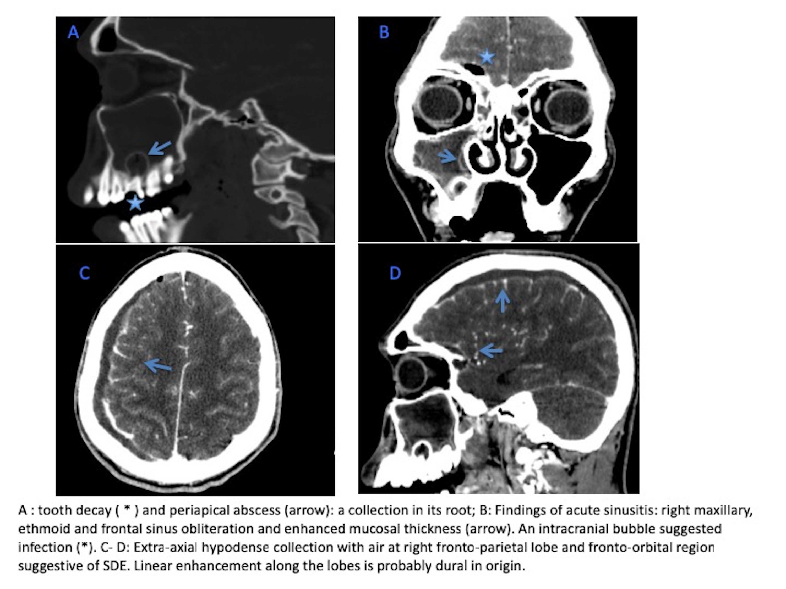
A 30-year-old male was referred from primary care due to worsening of acute sinusitis, with periorbital edema and intense right frontal headache. Enhanced- contrast sinus and head CT showed: - One superior right molar tooth with decay and a cavity filled with soft tissue content in its root that represents the periapical abscess. -The right maxillary and ethmoid sinus with enhanced mucosal thickness and sinus obliteration, Frontal sinus with air fluid levels. These findings are in keeping with acute sinusitis. An intracranial bubble adjacent to the frontal sinus suggested extension of the infection. - An extra-axial hypodense collection with air overlying the convexity of the right fronto-parietal lobe and fronto-orbital region suggestive of subdural empyema (SDE). Linear enhancement along the lobes is probably dural in origin. Diagnosis: Odontogenic sinusitis complicated with SDE. The patient was successfully treated with urgent surgical drainage of the subdural empyema and maxillary sinus, and molar exodontia.
Discusión
:Paranasal sinus infections are very common and dental abscesses are responsible for 10–12% of unilateral sinus infections. Most cases heal without complications. However sinusitis that is inadequately treated may lead to different complications: orbital (preseptal cellulitis, subperiosteal abscess), intracraneal (cavernous sinus thrombosis, meningitis, subdural empyema, epidural and intraparenchymal abscess). SDE although not frequent, is the most common intracranial complication in the setting of sinusitis. It may occur as a result of direct extension through the posterior frontal sinus, wall or through retrograde thrombophlebitis of the ophthalmic veins. It usually presents with a fulminant clinical course, and require prompt diagnosis and emergent neurosurgical drainage. Key points: - Considered head contrast-enhanced CT if the patient has acute sinusitis and neurologic symptoms. MRI is more sensitive and specific. CT may miss small collections. - Best diagnostic clue: extra-axial crescentic in shape hypodense collection with rim enhancement. - Differential diagnosis: - epidural empyema are typically lentiform (may coexist 15% of cases), - Chronic subdural hematoma: may be indistinguish, history may help, - subdural hygroma: no enhancing collection, often trauma history. - Sinus CT can provide valuable information regarding the cause of acute sinusitis, and should include coronal views with bone and soft tissue windows.
Conclusión
Odontogenic sinusitis can on rare occasions lead to serious, potentially lifethreatening intracranial complications. The radiologists must be able to make a precise and early diagnosis that may assure urgent aggressive medical and surgical management.
Bibliografía
- Osborn MK, Steinberg JP. Subdural empyema and other suppurative complications of paranasal sinusitis. Lancet Infect Dis. 2007,7: 62-67. - Osborn, A. Diagnostic Imaging: Brain. Philadelphia: Elsevier-USA, 2016. 654p.