Hospital: Hospital Universitario La Paz.
Nº: C2019-350
Aut@r o Autores: G. Moreno Montero, A. Díez Tascón, J. López, F. García, M. Caicoya, M. Martí De Gracia.
Presentación
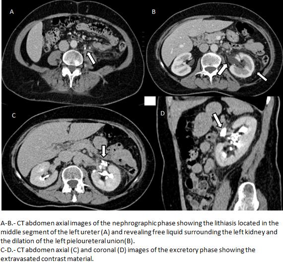
A 59-years old woman, with no relevant previous history, came to our hospital with a 4-hours history of left lumbar colic pain which radiates through her ipsilateral genital area, with no fever, nausea, vomits or urethral syndrome. At pshysical exploration, the abdomen was painful on the mesogastrium and left flank, with renal fist positive but Murphy and Blumberg both negative. Analytically, she had no pathologic values. US was made and we identified bilateral ectasy of the pieloureteral union and free liquid surrounding the left kidney. With these findings, a CT-urography with basal, nephrographic and excretory phases was completed. It was observed a left pieloureteral dilation secondary to a 4 mm obstructive lithiasis located in the mid segment of the left ureter, extravasated contrast material which seemed to originate from the rupture of the left medium anterior calyceal fornix and also left perirrenal free liquid.
Discusión
The findings are suggestive of a left obstructive uropathy with calyceal fornix rupture secondary to a lithiasis in the left ureter. Calyceal fornix is the weakest point of the renal collecting system, and its rupture is produced by an increase of the pressure in the excretory system. The most common etiology of renal forniceal rupture is obstruction caused by distal ureteric stones followed by malignant extrinsic ureteric compression. Regarding the symptomatology, emphasize that the patient’s symptoms may improve abruptly due to decompression caused by forniceal rupture. The diagnostic imaging of forniceal rupture is made based on the presence of any one of the criteria: irregularity of single renal calyx, loss of the ability to discern renal sinus fat, asymmetrically distributed perinephric stranding or a discrete perinephric fluid collection. The first study made is usually the US, and the CT is used to confirm the diagnosis. In the non-enhanced CT, the lithiasis is usually seen, and in theexcretory phase of the enhanced CT, the extravasated contrast material is identified surrounding the renal pelvis and proximal ureter. Initially, it is preferred medical treatment with or without double-J catheter. If there is infection or other complications, surgical treatment is the best option.
Conclusión
Forniceal rupture in an important pathology and it requires inmediate treatment. The most common etiology is obstruction caused by lithiasis and the CT is the best study to diagnose it.
Bibliografía
- Kawamoto S, Duggan P, Sheth S, Miyamoto H, Kazi Z, Fishman E. Renal Papillary and Calyceal Lesions at CT Urography: Genitourinary Imaging. RadioGraphics. 2017,37(1):358-9. - Kosehan D, Akin K, Topcu A, Koktener A, Cakir B, Teksam M. Spontaneous urinary