Hospital: Complejo Hospitalario Torrecárdenas.
Nº: C2019-671
Aut@r o Autores: V. Sánchez Miras, L. Zambrana Aguilar, J. García Díez.
Presentación
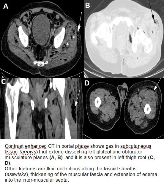
A 62-year-old male presented to emergency department with pain in left thigh, with purulent material draining and fever. He presented a clinical history of rectal squamous cell carcinoma 8 years earlier, treated with abdominoperineal resection and colostomy. After performing blood tests, the onset of sepsis was confirmed by high levels of CRP, lactic acid and leukocytosis. Abdominol-pelvic CT in venous portal phase shows the presence of gas in the subcutaneous tissue that extended from left perianal region to the left thigh root, dissecting obturator muscles and gluteal musculature. There are also ill-defined fluid collections along the deep fascial sheaths and signs of edema with extension into the inter-muscular septa. No intraabdominal pathological findings are observed. After these findings, the diagnosis of necrotizing fasciitis is established.
Discusión
Necrotizing fasciitis is a progressive, rapidly spreading infection of the deep fascia, with secondary necrosis of the subcutaneous tissues. The most common location are the extremities followed by the perineum, the trunk, and the head and neck. There are no true risk factors for necrotizing fasciitis, but there are predisposing factors such as injectable drug use, chronic debilitating comorbidities (eg, diabetes mellitus, immunosuppression, obesity) and peripheral vascular disease. Necrotizing soft-tissue infections are often accompanied by gas-forming anaerobic bacteria, usually in association with aerobic gram-negative organisms. The disease usually starts with inoculation of bacteria in the deep soft tissues after a skin wound. The main diagnostic dilemma is distinguishing celulitis from deep soft-tissue involvement occuring in necrotizing fascitis, which is more dangerous and requires more aggressive treatment, including surgical debridement. The main diagnostic sign that the radiology must look for is the presence of gas in the subcutaneous tissues caused by these anaerobic organisms, althoughgas is not always observed. Other CT features what should be considered include thickening of the affected fascia, fluid collections along the deep fascial sheaths and edema extension into the inter-muscular septa and muscles.
Conclusión
Necrotizing fasciitis constitutes a life-threatening surgical emergency. Unfortunately, this infection can be difficult to recognize in its early stages and it`s a rapidly progressive condition. Although establishing the diagnosis requires a high clinical suspicion, image studies can play a vital role the early diagnosis, allowing to perform an early and successful treatment.
Bibliografía
- Hayeri MR, Ziai P, Shehata ML, Teytelboym OM, Huang BK. Soft-Tissue Infections and Their Imaging Mimics: From Cellulitis to Necrotizing Fasciitis. RadioGraphics 2016, 36:1888–1910. - Malghem J, Lecouvet FE, Omoumi P, Maldague BE, Vande Berg BC. Necroti