Hospital: Hospital General de Albacete.
Nº: C2019-151
Aut@r o Autores: C. López Cárceles, I. Pérez Saus, A. Ibáñez Ibáñez, R. Rodenas Lozano, J. Rubio Medina, Á. Fernández López.
Presentación
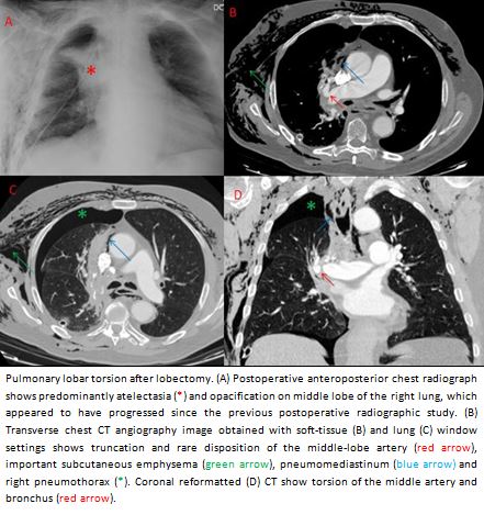
75-year-old man with adenocarcinoma of lung on rigth upper lobe. It was treated with surgery, perfonming a superior right lobectomy and mediastinal lymphadenectomy of the hilar, paratracheal and subcarinic groups by VATS (video assisted thoracic surgery), without incidents or immediate complications. 48 hours after the intervention, the patient presented important subcutaneous emphysema, chest X-ray it was also identified atelectasis of the middle lobe, not present in postoperative one. It was suspicion of torsion of the middle lobe by the findings of the radiograph, and chest CT angiography is performed, where the diagnosis is confirmed, and the patient was operated on urgently for detorsion.
Discusión
Lung torsion is the rotation of lung or lung lobe about its bronchovascular pedicle. It is a rare desease, that it presents a incidence of 0.09-0.4%. It can occur under three sets of circumstances: spontaneously, usually in association with some other pulmonary abnormality, following traumatic pneumothorax, most offen it occurs in the setting of lobectomy. The degree of pedicle rotation is variable, generally 180º, but on ocasion from 90° to 360°. It is an emergency, and it produces compromise of the airway, arterial blood supply that leads to lung ischemia, often shows hemorrhagic infarction or necrosis, and venous-lymphatic drainage resulting in interstitial edema and alveolar exudation. Radiorafhics features include at plain radiograph collapse or consolidated lobe, whith an unusual position, and CT findings include tapered obliteration of the proximal pulmonary artery and accompanying bronchus of the involved lobe and amorphous soft-tissue attenuation at the hilum, whith poorly enhancing consolidation of the lung and ground-glass attenuation. CT angiography with a automated bolus detection system (SmartPrep) in main pulmonary artery could be used. Treatement is surgical fixation, and mortality rate is very high if the torsion goes unrecognized.
Conclusión
Pulmonary torsion is a rare disease and a rare complication of thoracic surgeries, but it is a medical emergency with a high mortality rate, which is why we must know it, in order to diagnose it, because early recognition is vital, and CT angiography may be helpful in making an early confirmatory diagnosis
Bibliografía
- Radiographic and CT findings in complications following pulmonary resection. Kim EA, Lee KS, Shim YM, Kim J, Kim K, Kim TS, Yang PS. Radiographics. 2002 Jan-Feb,22(1):67-86. - Emergent and nonemergent nonbowel torsion: spectrum of imaging and clinical f