Hospital: Hospital Clínico Universitario Virgen de la Arrixaca.
Nº: C2019-432
Aut@r o Autores: I. Sánchez-Serrano, A. Cuélliga González, A. León Hernández, A. Navarro Baño, M. Martínez Cutillas, M. Ato González.
Presentación
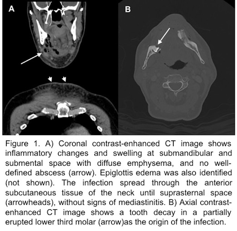
65-years-old man presented to the emergency department with pain and enlargement of the floor of the mouth and difficulty to breath and speak. Physical examination revealed an increase of soft tissue at the submental and submandibular space. Analytics showed a rise of the acute phase reactants and leukocytosis. A contrast-enhanced CT revealed inflammatory changes and swelling at submandibular and submental space with diffuse emphysema, and no well-defined abscess, associated with inflammation of the anterior subcutaneous tissue of the neck until suprasternal space (Figure 1). A tooth decay was found in a partially erupted lower third molar (piece 48). The diagnosis of Ludwig’s angina secondary to dental infection was proposed and the patient was undergoing surgical treatment.
Discusión
Ludwig’s angina is a life-threatening cellulitis of the floor of the mouth. It usually arises from a tooth infection, most often the third mandibular molar, and is caused by multiples microorganism, even gas-forming bacteria. Although it may debut as an edema of the mouth floor, it may also present emphysema in the context of a necrotizing soft-tissue infection. If a correct management is not performed, this infection can be lethal, due to the fast spread to the mediastinum and the compromise of the airway [1, 2].
Conclusión
Correct radiologic diagnosis of angina de Ludwig is essential because of the severity of this pathology, and in order to avoid life-threatening complications like mediastinitis or airway compromise.
Bibliografía
- Capps EF, Kinsella JJ, Gupta M, Bhatki AM, Opatowsky MJ. Emergency imaging assessment of acute, nontraumatic conditions of the head and neck. Radiographics. 2010,30(5):1335-52. doi: 10.1148/rg.305105040 - Reynolds SC, Chow AW. Life-threatening infectio