Hospital: Corporació Sanitària Parc Taulí.
Nº: C2019-425
Aut@r o Autores: P. Escarcena, B. Consola.
Presentación
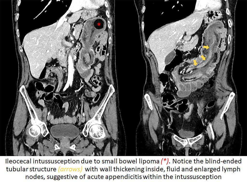
A 53-year-old woman was admitted to the emergency department with a threeday history of abdominal pain refractory to analgesics, she had no fever, vomiting, ot other symptoms. She had a lipomatous mass in the ileocecal valve diagnosed 10 years before and a history of recurrent episodes of intussusception. The latest follow-up showed stable disease. Laboratory test revealed elevated WBC count (12150/mL) and C-reactive protein (17.68 mg/L). Abdominal CT for clinical suspicion of intestinal intussusception confirmed progression of the ileocecal intussusception and identified a blind-ended tubular structure with wall thickening inside, suggestive of acute appendicitis within the intussusception, as well as a slight amount of fluid, stranding of the adjacent fat and enlarged lymph nodes within the intussusception. Surgery confirmed the ileocecal intussusception and inflamed appendix within it. Histopathologic examination of the resected appendix showed signs of acute phlegmonous inflammation and periappendicitis.
Discusión
The clinical manifestations of intussusception and acute appendicitis can be similar, including abdominal pain and vomiting. Although in adults acute appendicitis is much more common than intussusception, it is easy to overlook on of these conditions when we suspect the other. Therefore, it is important to consider the possibility that both conditions may coexist. If undiagnosed in the early stages, both conditions can lead to severe complications such as peritonitis and death. We present an interesting case of acute appendicitis within an ileocolic intussusception, a very rare phenomenon. In our case, the diagnosis of an intussuscepted appendix with signs of inflammation was suggested at imaging and then confirmed on histopathologic examination of the surgical specimen. However, in some series, only about on.third of cases were diagnosed preoperatively, with the remaining cases diagnosed through surgery. These results show the challenge radiologists face in diagnosing this coindition and the importance of familiarity with the imaging features.
Conclusión
Although rare, acute appendicitis within an intussusception should be considered when evaluating patients with suspected ileocecal intussuscepction.
Bibliografía
- Kim YH, Blake MA, Harisinghani MG, Archer-Arroyo K, Hahn PF, Pitman MB, et al. Adult intestinal intussusception: CT appearances and identification of a causative lead point. Radiographics 2006, 26(3):733-44. - Luzier J, Verhey P, Dobos N. Preoperative