Hospital: Complejo Asistencial Universitario de Salamanca.
Nº: C2019-645
Aut@r o Autores: D. Vargas Jiménez, J. Santos Sanchez, M. Coderque Mejía, M. Ibarra Hernández, S. Márquez Batalla, J. Uzcátegui León.
Presentación
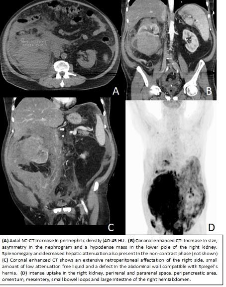
We report a case of a 68-year-old without relevant medical history with a complaint of two days of dyspnea, abdominal pain and macroscopic hematuria. At the physical examination, he was tachycardic, tachypneic, and hypotensive. The abdominal palpation revealed a mass on the right flank. Laboratory tests showed an increase in creatinine, arterial lactate, acute phase reactants, lactate dehydrogenase, the blood count showed normocytic normochromic anemia with normal leukocytes. An urgent non-contrast abdominal CT and a nephrographic and excretory phase after the administration of iodinated contrast was performed. In non-contrast CT (NCCT) an increase in retroperitoneal density (40-45 HU) was observed mainly in the perirenal and right pararenal associated with size increase of the ipsilateral kidney. In the nephrographic phase, the extensive retroperitoneal affectation of the right side, increase in size, asymmetry in the nephrogram and a hypodense mass in the lower pole was confirmed, in the excretory phase there was an absent elimination of the right kidney. Due to radiological suspicion of shattered kidney with retroperitoneal hematoma and sustained hypotension an exploratory laparotomy was performed. A transperitoneal subcostal approach was completed discarding the possibility of bleeding in the abdominal cavity. In surgery, a hard and unresectable mass and a small amount of non-blood fluid was found. Samples of this mass, omentum and fluid were sent for anatomopathological analysis. The analyzes found renal and peritoneal infiltration by diffuse large B-cell lymphoma (DLBCL). An FDG PET- CT was performed demonstrating an intense uptake in the right kidney, perirenal and pararenal space, peripancreatic area, omentum, mesentery, small bowel loops and large intestine of the right hemiabdomen. These findings are compatible with stage IV lymphoma.
Discusión
Renal involvement by lymphoma it’s most common in patients with non-Hodgkin disease, especially DLBCL with advanced-stage extranodal disease. The clinical presentation is often, pain, weight loss, hematuria and palpable mass. Multiple patterns of lymphomatous involvement have been described. Although the perirenal extension from the retroperitoneum or from the kidney due transcapsular spread is not infrequent, it’s probably the most atypical and pathognomonic. Consists of a tissue plate that surrounds the kidney, usually, the tissue is iso hyperdense with respect to renal parenchyma in NCCT and hypodense after contrast.
Conclusión
Renal involvement by lymphoma it’s most common in advanced-stage extranodal disease, due iso-hyperdensity of this affectation, hematomas must be included as a differential diagnosis, the extent of local neoplasms of the retroperitoneal viscera, other inflammatory processes such as urinomas, pancreatitis or peritoneal fibrosis must be included too.
Bibliografía
- Purysko A et al. Imaging Manifestations of Hematologic Diseases with Renal and Perinephric Involvement. RadioGraphics 2016, 36:1038–1054. - Sheet S, Ali S, Fishman E. Imaging of Renal Lymphoma: Patterns of Disease with Pathologic Correlation. RadioGrap