Hospital: HOSPITAL UNIVERSITARIO DE ALAVA.
Nº: C2019-428
Aut@r o Autores: N. Serrano Usaola, S. Beltran De Otalora Garcia, M. Martin Egaña, L. Alonso Irigaray, R. Gonzalez Serrano, D. Del Pozo Alcorta.
Presentación
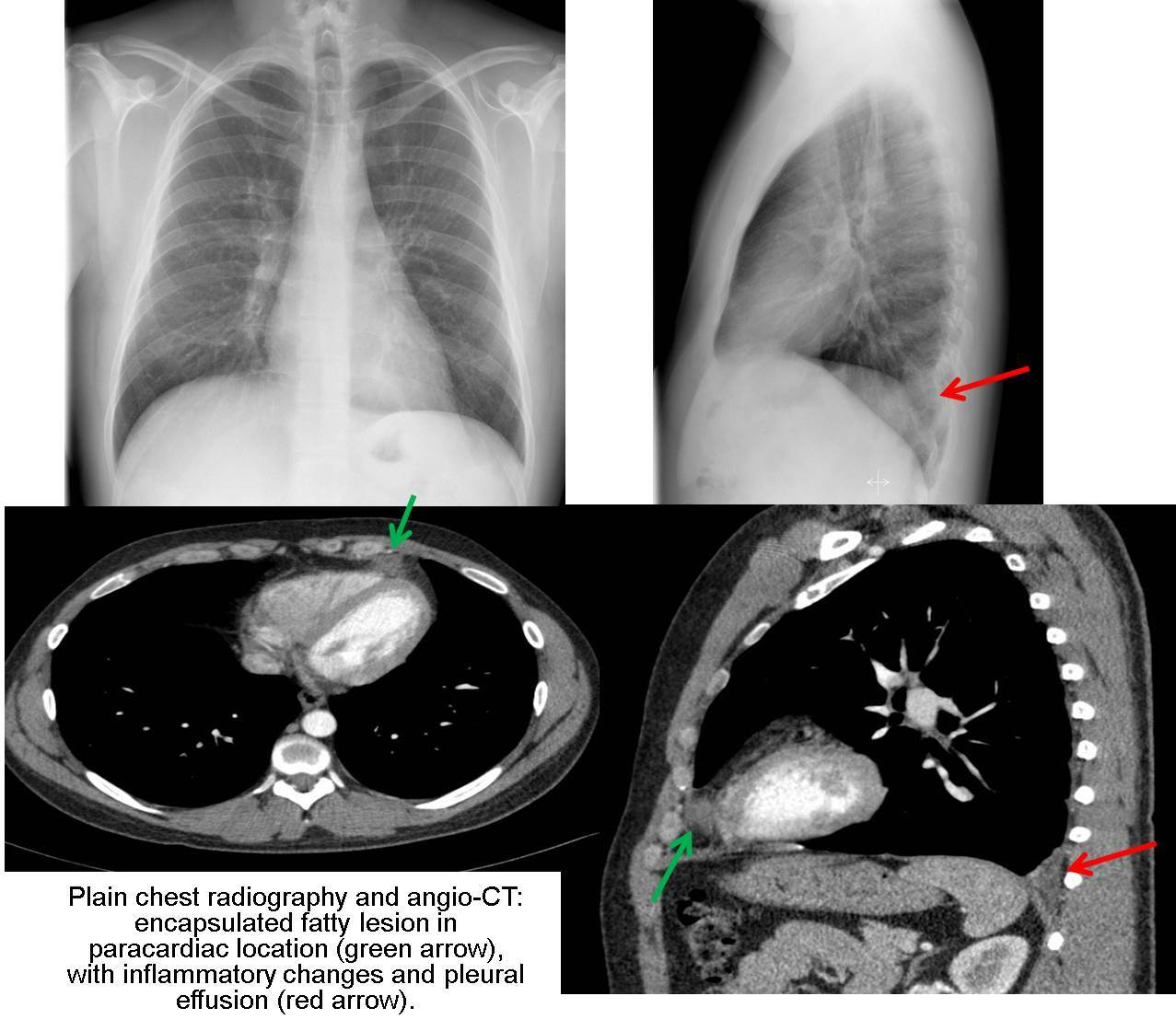
A 35 year-old man came to the emergency room of our hospital, with acute pleuritic chest pain and respiratory insufficiency. He had no previous medical history. Physical examination, laboratory tests and ECG were normal. In plain chest radiography, AP and lateral, a posterior left costophrenic sinus impingement was noted, probably the expression of a little pleural effusion. A CT-angiogram of the pulmonary vasculature was performed to exclude a pulmonary embolism, which was excluded, and revealed a high attenuation lesion in the epi-pericardial fat with central hipodensity and encapsulated form, with inflammatory changes in the nearby fat and pleural effusion, already detected in the X-ray. Based on clinical and radiological findings, the diagnosis of epi-pericardial fat necrosis was performed. Patient was treated with analgesics, with satisfactory clinical evolution and without needing for more aggressive procedures.
Discusión
Epi-pericardial fat necrosis is an uncommon disorder, with sefl-limiting and bening course. Its etiology is unknown and pathologic findings are similar to those found in fat necrosis of epiploic appendagitis, for example. The clinical presentation consists of acute and intense pleuritic pain, in a previously asymptomatic and healthy patient. It may associate dyspnea, syncope...mimicking the symptoms of myocardial infarctation and/or pulmonary thromboembolism. Physical examination, laboratory tests and ECG are, usually normal. In imaging diagnosis, plain chest X-ray may be normal or show paracardiac opacity, usually left side, with or without associated pleural effusion. The CT, considered the image technique of choice for the diagnosis in case of suspicion, will reveal an encapsulated fatty lesion in paracardiac location, with inflammatory changes, pericardial and/or pleural thickening and occasionally pleural effusion. In doubtful or inconclusive cases on CT, MRI can confirm the fat content of the lesion. The differential diagnosis of this entity should be made, based on the clinical presentation with pulmonary embolism and acute coronary disease, because for its frequency, severity and prognosis, and based on the radiological findings with fatty lesions of paracardiac location (lipoma, liposarcomas...), diaphragmatic hernias occupying the cardiofrenic space, or others. Taking into account the self-limited and benign course of this entity, the management is conservative, with analgesic and antiinflammatory drugs, for pain control. Radiologicalcontrol, with CT, can be performed in 1-2 months, which will show the resolution of the findings.
Conclusión
Epi-pericardial fat necrosis is an uncommon entity, with benign and self-limiting course, that must be considered in the diagnosis of patient with acute thoracic pain, previously asymptomatic and healthy. The combination of characteristic clinical and radiological findings allows to establish the diagnosis and avoid more aggressive procedures.
Bibliografía
- Pineda V, Andreu J, Cáceres J, Merino X, Varona D, Dominguez-Oronoz R. Lesions of the cardiophrenic space: findings at crosssectional imaging. Radiographics 2007,27:1932 - Pineda V, Cáceres J, Andreu J, Vilar J, Domingo ML. Epipericardial