Hospital: Hospital Universitario San Cecilio, Hospital Santa Ana.
Nº: C2019-201
Aut@r o Autores: L. Díaz Rubia, Y. Núñez Delgado, P. García-Villanova Ruíz, L. Guirado Isla, F. García Verdejo.
Presentación
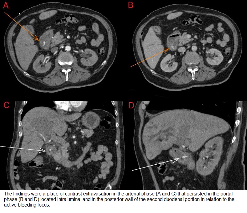
This is a 75-year-old man with a history of HCV and hepatocellular carcinoma treated with chemoembolization that was admited to the hospital due to liver failure, renal failure and anemia with 9 g/dl of hemoglobin. On the second day of hopitalization, he suffered sudden deterioration of the general condition along with melena and anemization with hemoglobin of 4 g/dl, so it was decided to request a CT angiography of the abdomen. A basal study without contrast was performed, an arterial phase with bolus tracking technique with infradiaphragmatic aortic region of interes and a portal phase. The findings were a place of contrast extravasation in the arterial phase that persisted in the portal phase located intraluminal and in the posterior wall of the second duodenal portion in relation to the active bleeding focus (figure 1). After this, it was decided to perform emergency upper gastrointestinal endoscopy in which an ulcerated type Forrest III lesion was detected in the second duodenal portion with abundant bleeding treated with sclerosis with adrenaline and hemoclips. The patient evolved favorably and could be discharged in the following days.
Discusión
Bleeding of the upper gastrointestinal tract is caused by erosion or ulceration in up to 55-75% of cases, and less frequent by varicose veins, vascular lesions or neoplasms. Its diagnosis by CT angiography has a high sensitivity and specificity and constitutes a challenge for the radiologist since sometimes it is not possible to locate the active bleeding point and only indirect data of hemorrhage can be seen. It is very important to perform a first study without contrast in which we will assess the existence or not of dense content in the intestinal lumen, the presence of hematomas, etc. In the arterial phase study, active bleeding can be seen as a source of contrast extravasation in the affected bowel lumen, and it is sometimes necessary to perform venous and late phases to ensure diagnosis.
Conclusión
CT angiography is a very useful noninvasive exploration for the diagnosis of intestinal active bleeding. Among its advantages, it stands out its rapidity of execution, a post-processing that is not very laborious and high availability, which will allow a diagnosis and an early treatment that improves the prognosis of these patients.
Bibliografía
- Geffroy Y, Rodallec MH, Boulay-Coletta I, Jullès MC, Ridereau-Zins M. Multidetector CT angiography in acute gastrointestinal bleeding : why , when and how. Radiographics 2011 May-Jun,31(3):E35-46. - Lee SS, Oh TS, Kim HJ, Chung JW, Park SH, Kim AY, et