Hospital: Hospital Universitario la Paz.
Nº: C2019-577
Aut@r o Autores: J.M. López Vega, C. Oterino Serrano, G. Buitrago Weiland, A. Diez Tascon, M. Martí Degracia.
Presentación
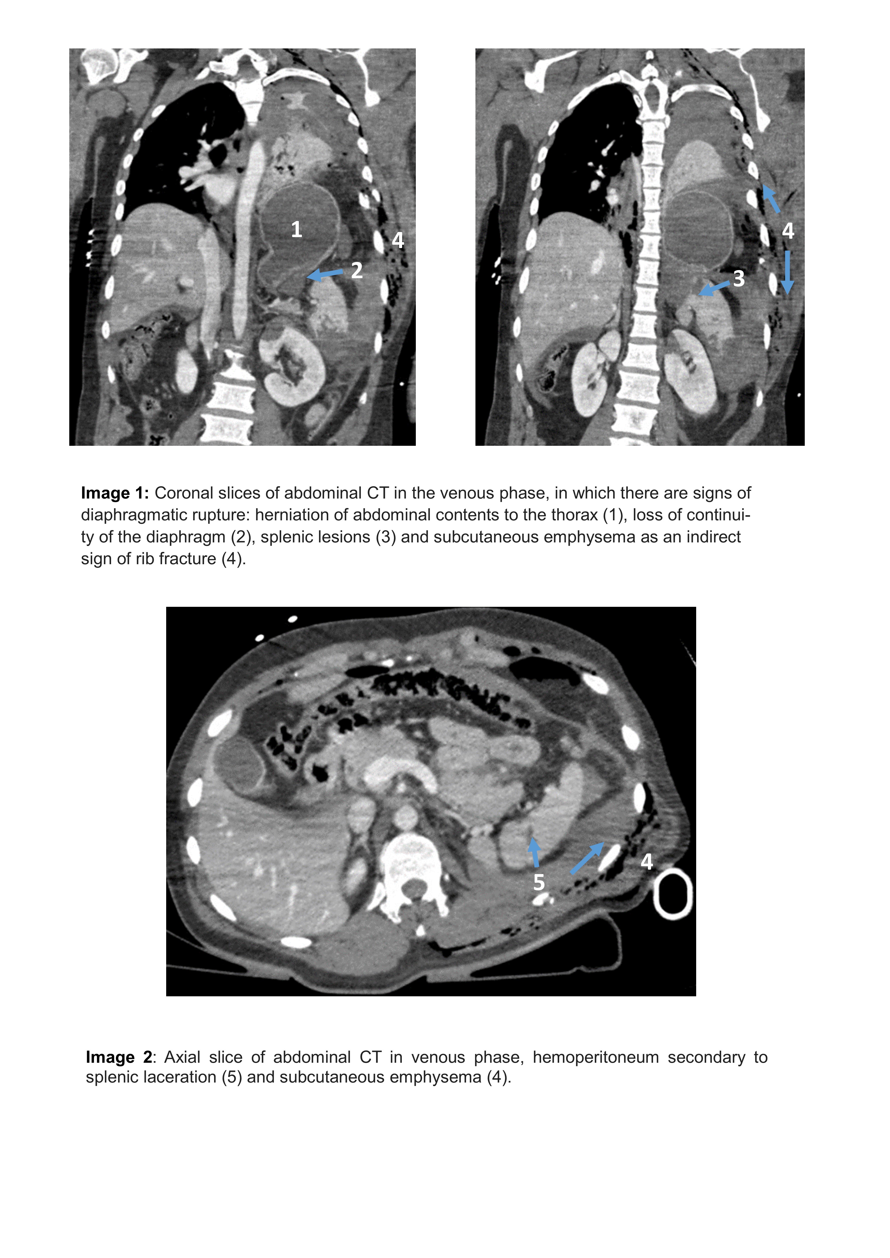
We present the case of a 42-year-old man who suffered a work trauma due to precipitation from 6 meters high. He presented with a decrease in the level of consciousness and hemodynamic instability. In the hospitalary FAST free fluid was identified. Afterwards, a body CT was performed, showing intrathoracic herniation of the stomach, the splenic angle and the transverse colon with a wide continuity solution in the left hemidiaphragm. These findings suggest traumatic diaphragmatic rupture with herniation. Emergency surgical repair of the lesions was performed with satisfactory patient recovery.
Discusión
The diaphragmatic rupture is a loss of continuity of the muscle fibers that leads to a communication between the abdominal and thoracic cavity. Given the severity of this case, abdominal CT allows a reliable diagnosis of diaphragmatic rupture since herniation over the defect is seen, being this the technique of initial choice, preferring it over other techniques such as ultrasound or plain radiography. In the CT other radiological signs that help to guide a diaphragmatic rupture, when herniation is not estimable, making special emphasis on the left side given the position of the liver that usually prevents herniation, include: a segmental diaphragmatic defect due to loss of continuity, focal thickening of the hemidiaphragm, absence of part of the diaphragm, the presence of a hump sign, hypoenhanced diaphragm, extravasation of contrast, associated lesions of other organs and, in some occasions, important vascular traumas. The observation of two or more of those CT signs is associated with a substantially higher probability of the presence of diaphragmatic rupture. These signs may alert the radiologist to the need to examine the diaphragm more closely for other features supportive of the diagnosis or its exclusion. For patients with an uncertain diagnosis after CT, MR imaging should be performed.
Conclusión
In conclusion, diaphragmatic rupture should be suspected in patients who suffer blunt thoracoabdominal high-impact trauma. Their study should be conducted through the use of CT focusing especially on the left side of the diaphragm. Although mortality is relatively low, the risk of complications persists throughout life due to the diaphragm is rarely repaired spontaneously, so timely diagnosis allows early treatment.
Bibliografía
- Iochum S, Ludig T, Walter F, Sebbag H, Grosdidier G, Blum AG. Imaging of Diaphragmatic Injury?: A Diagnostic Challenge?? Radio Graph. 2002,22:103–16. - Eren S, Kantarci M, Okur A. Imaging of diaphragmatic rupture after trauma. Clin Radiol. 2006,61(6)