Hospital: Hospital Universitario Ramón y Cajal.
Nº: C2019-322
Aut@r o Autores: J. Blanc Molina, A. Silva Rodríguez, B. Alba Pérez, A. López-Frías López-Jurado, P. Marazuela García, C. Suevos Ballesteros.
Presentación
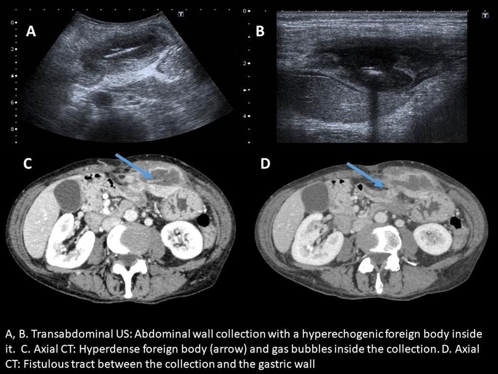
A 62 year-old woman was admitted to our Emergency Department with abdominal pain that started 15 days ago and a mesogastrium palpable abdominal mass. She has no fever and laboratory tests revealed increased acute phase reactants.Abdominal US and CT were performed. They showed a well-defined fluid collection with hypercaptant walls on CT in the left rectus abdominis muscle. It was in intimate contact with the gastric wall, which it was thinned and edematous, and a fistulous tract between them was identified. Inside the collection there was a lineal image that showed slightly hyperdense on CT and hyperechogenic with posterior acoustic shadowing on US, and isolated gas bubbles. There was no pneumoperitoneum or ascites.
Discusión
Our diagnosis was a contained gastric perforation due to a foreign body (probably a fish bone). The patient went through surgery, where the collection, with purulent material, was drained and the foreign body was found but, unfortunately, not identified.
Conclusión
Foreign body perforation of the gastrointestinal tract has a wide spectrum of clinical presentation and sometimes the symptoms are subacute or chronic. The perforation is caused by progressive erosion of the foreign body, so free intraperitoneal air is not usually seen.
Bibliografía
-Gayer G, Petrovitch I, Jeffrey R. Foreign Objects Encountered in the Abdominal Cavity at CT. RadioGraphics. 2011,31(2):409-428. - Goh B, Tan Y, Lin S, Chow P, Cheah F, Ooi L et al. CT in the Preoperative Diagnosis of Fish Bone Perforation of the Gastro