Hospital: HOSPITAL UNIVERSITARIO BASURTO.
Nº: C2019-243
Aut@r o Autores: M. Santamaria, O. Gorriño, A. Legorburu, I. Lecumberri, J. Gomez.
Presentación
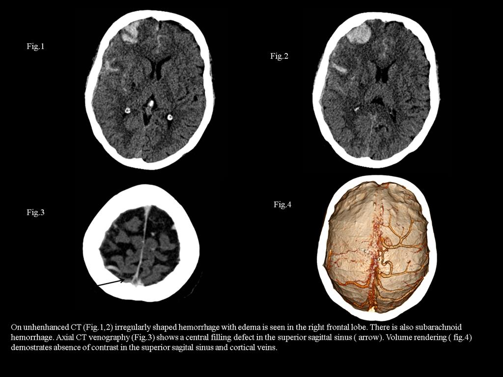
53-year-old woman with severe headache with a history of breast cancer and hormonal treatment. Unenhanced CT and CT venography are performed at the emergency department. On unenhanced CT irregularly shaped cortico-subcortical hemorrhage with edema is seen in the right frontal lobe. There is also subarachnoid hemorrhage. Axial CT venography shows a central filling defect in the superior sagittal sinus. Volume rendering demonstrates absence of contrast in the superior sagittal sinus and cortical veins.
Discusión
Cerebral venous thrombosis is a rare neurologic disorder potentially reversible with a prompt and appropriate diagnosis and medical therapy. Imaging plays a primary role in the diagnosis because causal factors and clinical manifestations are many and varied. Many causes have been described and can be classified as local (related to cerebral veins and dural sinuses) or systemic (related to conditions that promote thrombosis). Clinical presentation is often nonspecific. Common symptoms and signs include headache, papilledema, seizures, focal neurological deficits and mental status change. Obstruction of venous drainage with increasing venous pressure in the affected region of brain produces extravasation of fluid. If venous pressure continues to increase there is a consequent diminishment in arterial perfusion pressure and cell death may ensue. Thus, parenchymal changes may be secondary to cytotoxic edema, vasogenic edema or hemorrhage. CT is often the first imaging modality performed at the emergency department. In patients with unenhanced CT findings suggestive of venous thrombosis, CT venography can be performed without delay to confirm the diagnosis and to start appropriate therapy immediately. Direct signs are the dense sinus and the delta sign on enhanced CT. Indirect signs are the edema and cortical hemorrhage. Both CTV and MRI are complementary and may be combined for optimal evaluation in complex or equivocal cases. MRI enables better delineation of parenchymal abnormalities compared to CT.
Conclusión
Key point Diagnosis of cerebral venous thrombosis is challenging. Prompt diagnosis can lead to effective treatment and reduce morbidity and mortality. CT venography can be performed at emergency department if unenhanced CT suggests venous thrombosis. Knowledge of the normal anatomy of the intracranial venous system and the most frequent variations is useful for accurate interpretation of venographic images and identification of the drainage territories. Recognition of patterns of parenchimal abnormalities and hemorrhage compared with arterial infarcts is key for early recognition and work-up of cerebral venous thrombosis
Bibliografía
- Leach JL, Fortuna RB, Jones BV, Gaskill-Shipley MF. Imaging of Cerebral Venous Thrombosis: Current Techniques, Spectrum of Findings and Diagnostic Pitfalls. Radiographics 2006,26:S19-S43. - Rodallec MH, Krainik A, Feydy A, Helias A, Colombani JM, Julle