Hospital: Hospital Universitario Central de Asturias.
Nº: C2019-265
Aut@r o Autores: R. Viveros Vargas, N. Sordo Alonso, D. García Pérez, D. Vizcaíno Domínguez, M. Tijerín Bueno, J. Calvo Blanco.
Presentación
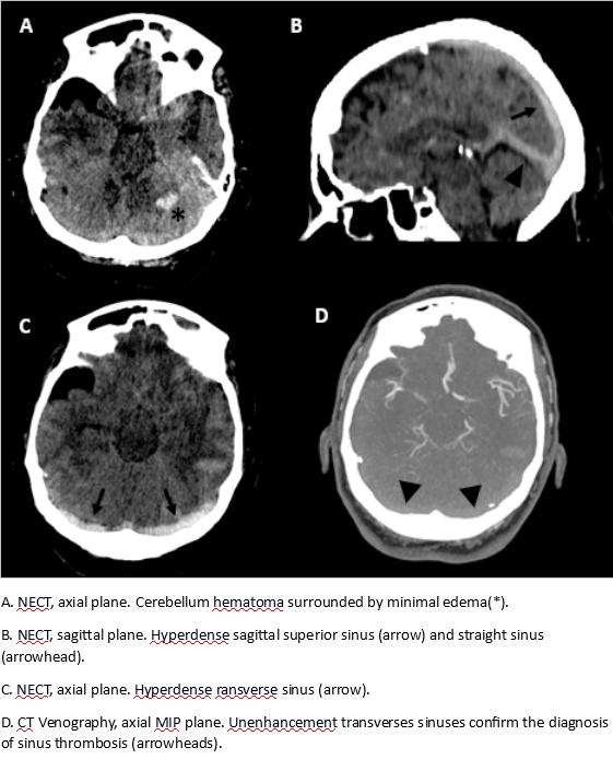
A 50-year-old woman is referred to Emergency Department by her primary care physician for intense frontal headache and vomiting. Physical exploration reveals a low level of consciousness and Glasgow score 10. Cerebral CT is required. FINDINGS: Intraparenchymal haematoma in the left cerebellar hemisphere with minimal edema. Hyperdensity of the transverse sinuses, straight and superior sagittal sinuses. CT venography is performanced demonstrating unenhancement of venous sinuses.
Discusión
DiagnosIs: Venous infarction secondary to venous sinus thrombosis. Cerebral venous sinus thrombosis is an uncommon cerebrovascular event, accounting for 0.5%– 1% of cases of stroke. This a young adults disease and its diagnosis is based on clinical suspicion and neuroimaging. An early diagnosis is important in order to early anticoagulation in thought to prevent thrombus propagation. As most patients present nonspecific symptoms, nonenhanced CT is the initial technique of imaging. Nonenhanced CT finding: -NECT is reported as normal in 2/3 of cases. -The findings in venous sinus thrombosis in early stages are hyperdense venous sinus and cerebral swelling. Hyperdense venous sinus (dense triangle or cord sign) is a sensitive sign for cerebral venous sinus thrombosis. -Venous infarcts and fragmented haemorrhage are late signs and are seen as low-attenuation lesions with or without subcortical haemorrhage. Subarachnoid haemorrhage of convexity is a rare manifestation. As soon as a suspicion of sinus thrombosis is established, the next technique is CT venography. CT venography findings and CECT: -Not enhancement of sinus or localized filling defect. -Empty delta sign (dura enhanced without trombus enhancement). -Filling defects should not be confused with Pacchionian bodies (arachnoid granulations).
Conclusión
-Cerebral venous sinus thrombosis is an uncommon cerebrovascular event. It is a diagnostic challenge for the clinical physician and the radiologist because of unspecify of clinical and radiological findings in early stage. -2/3 of nonenhanced CT in patien
Bibliografía
- Rodallec MH, Krainik A, Feydy A, et al. Cerebral Venous Thrombosis and Multidetector CT Angiography: Tips and Tricks. RadioGraphics 2006,26:S5-S18 - Herrmann KA, Sporer B, Yousry TA. Thrombosis of the internal cerebral vein associated with transient uni