Hospital: Hospital Universitario la Paz.
Nº: C2019-665
Aut@r o Autores: P. Carreño Moran, S. Yañez Castaño, D. Vargas Jimenez, P. Arias Rodriguez, A. Sanchez Martin, J. Uzcategui Leon. CAUSA Complejo Asistencial Salamanca,
Presentación
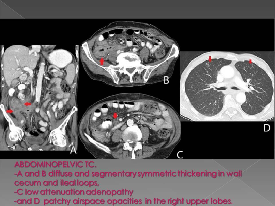
73 years hepatic transplantation, constitutional syndrome and immunosuppressive therapy. Colonoscopy: ulcerated lesion in cecum. Abdominopelvic TC: diffuse and segmentary symmetric thickening in wall cecum and ileal loops, low attenuation adenopathy and patchy airspace opacities in the right upper lobes.?
Discusión
DIFERENCIAL DIAGNOSIS ALGORITHMIC: SPECTRUM OF FIVEATTENUATION PATTERNS OBSERVED IN BOWEL WALL DISEASE: 1. Bowell wall thickening. a) focal 40cm suggest Benign, (except lymphoma 3 patterns: aneurismatic dilated wall, polypoid o adenopathy’s). 2. Pattern of attenuation: -White: ischemia or active inflammatory bowel disease (IBD). Avid contrast material enhancement that uniformly affects the majority of the thickened bowel wall. Suggest 2 pathologies: (a) vasodilation and/or (b) injury to intramural vessels with accompanying interstitial leakage. “Shock bowel” diffuse ischemia of the small bowel in hypotensive adults who have sustained blunt trauma) white attenuation pattern represents a reversible ischemic. -Gray: ischemia, chronic IBD o radiation enteritis (RE) Stratified: ischemia/vasculitis, active IBD, infection, RE, portal hypertension - Black: pneumatosis ischemic (portal o vein gas), infection o benign. - Water halo: stratification within a thickened bowel wall that consists of either 2 o 3 (“target sign”: ) continuous, symmetrically thickened layers. Halo sign with two layers (double halo) is composed of either a higher-attenuation outer annular ring surrounding a second, is most valuable as an unequivocal observation of bowel wall injury, often of an acute nature. Infections, IBD, vascular disease o RE - The pattern of the fat halo sign refers to a three-layered target sign of thickened bowel in which the middle or “submucosal” layer has a fatty attenuation (<10UH) : colitis ulcerosa, RE .Diferencial diagnosis: - Mycobacteria’s - Sarcoidosis - Lymphoma
Conclusión
- Mycobacterium tuberculosis positive. Diagnosis: TBC cecoileitis. - Inmunosupresión, QTP, o immunotherapy : high risk reactivation TBC - Differential diagnosis algorithm is an useful tool.26.
Bibliografía
- Sugi MD, Menias CO, Lubner MG, Bhalla S, Mellnick VM, Kwon MH, Katz DS. CT Findings of Acute Small-Bowell Entities. Radiographics. 2018 , 38: 13521369. - Fernandes T, Oliveira MI, Castro R, Araújo B, Viamonte B, Cunha R. Bowel wall thickening at