Hospital: H.U. Puerta de Hierro.
Nº: C2019-371
Aut@r o Autores: M. Reyero Lafuente, A. Sánchez Ramos, M. Collado Torres, E. Van Den Brule Rodríguez De Medina, A. Alcolado Jaramillo, M.A. Pastrana Ledesma.
Presentación
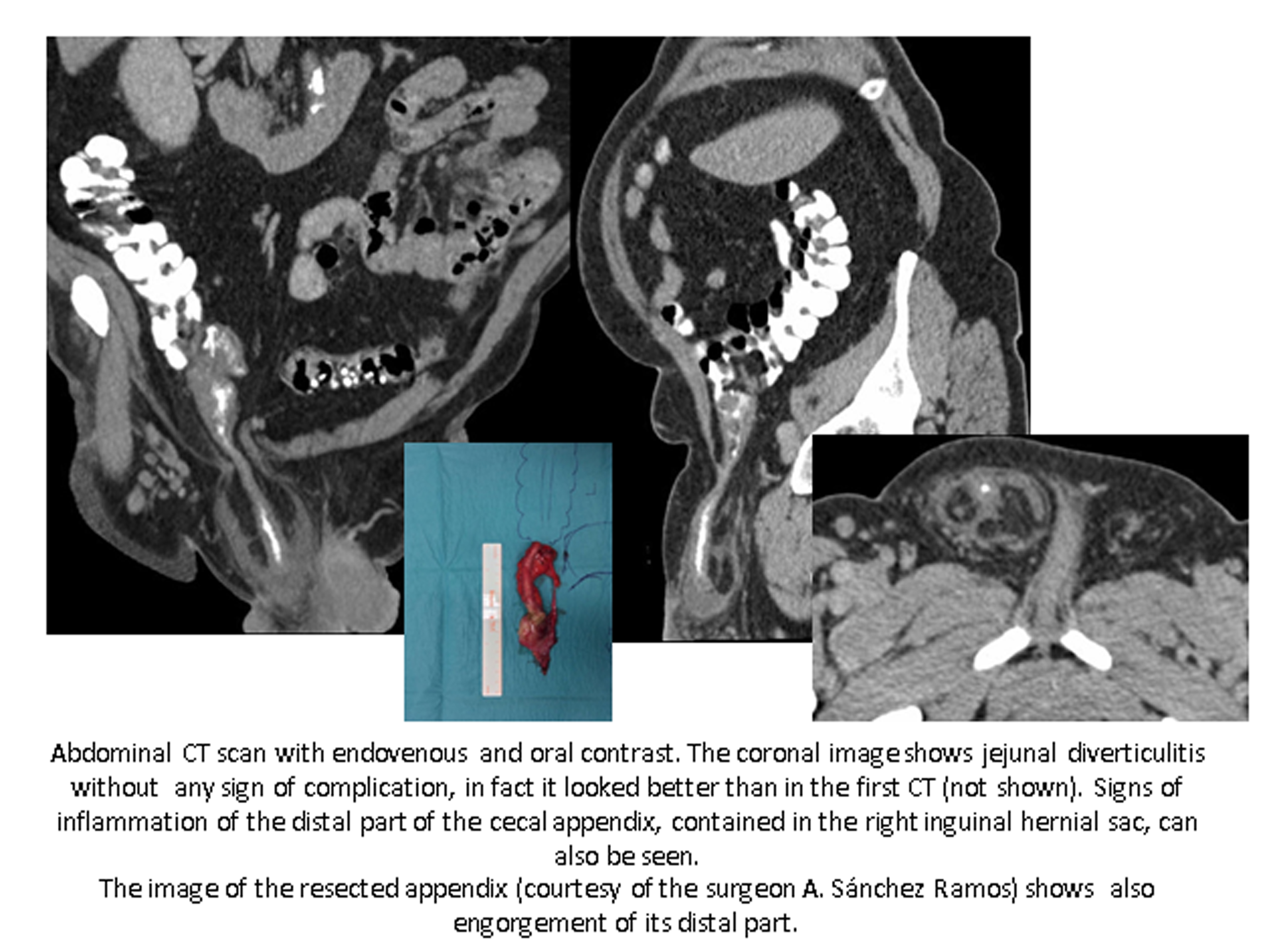
We report the case of a 65-year-old male complaining about persistent abdominal pain during hospitalization for conservative treatment due to jejunal diverticulitis. Abdominal tenderness with no signs of peritoneal irritation was observed as well as a non-complicated right inguinal hernia. The blood test showed leukocytosis and C-reactive-protein high levels. No signs of bowel obstruction were seen on the abdominal X-ray. Abdominal CT with oral and intravenous contrast revealed no signs of jejunal diverticulitis worsening or complication. Instead of it, acute inflammation signs of the cecal appendix which was surprisingly located in the right inguinal hernia were found. Urgent laparoscopic abdominal exploration was carried out confirming radiological findings and appendectomy was performed. Furthermore, right inguinal hernioplasty was held using an anterior open approach, in order to avoid mesh deployment in a contaminated surgical site.
Discusión
Despite finding inflammation typical signs, the real challenge, in this case, is locating the appendix. Usually situated in the right lower abdomen, the appendix may be rarely found in other locations such as left lower abdomen, in patients with intestinal malrotation, femoral canal (De Garengeot hernia) or inguinal canal (Amyand hernia). To find the appendix it is mandatory looking for the cecum first. This method should be used in all the radiologic techniques.
Conclusión
Amyand’s hernia is a rare form of inguinal hernia defined by the containing of the appendix. Incarceration is its most frequent presentation while inflammation of its content is more unlikely as well as accomplishing its clinical suspicion. We present a case of an atypical location of the cecal appendix with an infrequent complication.
Bibliografía
- Chin C.M., Lim K.L. Appendicitis: Atypical and challenging CT appearances: Resident and fellow education feature. Radiographics, 2015, Vol. 35, Issue 1 - Cabarrus M.C., Yeh B.M., Phelps A.S., Ou J.J., Behr S.C. From inguinal hernias to spermatic cord