Hospital: Hospital Universitario Virgen de la Arrixaca.
Nº: C2019-382
Aut@r o Autores: S. Ibañez-Caturla, A. Cuélliga-González, G. Litrán-López, A. Jiménez-Sánchez, F. Sarabia-Tirado, M. Carrillo-García.
Presentación
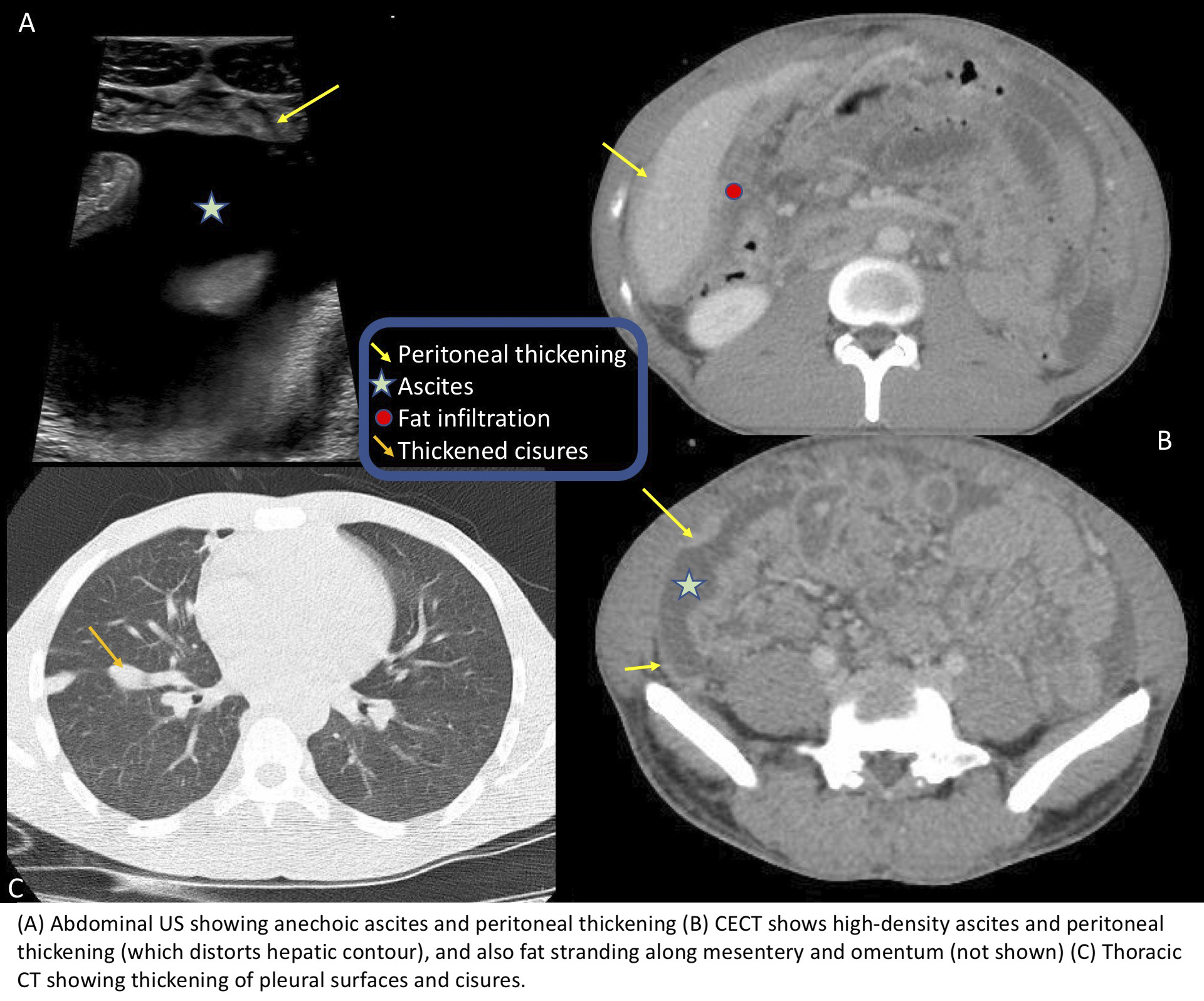
32-year-old male born in Gambia, attending the emergency department due to continuous diffuse abdominal pain that started one week earlier, several episodes of diarrhea, low-grade fever and chills. He refers undetermined weight loss in the last month. No further symptoms. Blood pressure and heart rate were normal. Physical exploration was quite unremarkable. Blood tests showed increased C-reactive protein and white-cell count. An abdominal ultrasound was requested, which showed focal peritoneal thickening, distributed throughout the abdominal cavity and producing mass effect over hepatic and splenic parenchyma, and also moderate amount of ascites. Abdominal CT scan also revealed thickened and enhanced peritoneal lining, with hyperattenuating ascites and increased density and micronodules over omental fat. No lymph node enlargement or other signs of malignancy. Pulmonary bases showed nodular thickening of the pleural and cisural linings, and therefore a thoracic CT was performed, which also showed a small consolidation and micronodular tree-in-bud opacities on the anterior upper-right lobe. All these findings were suggestive of pleural and peritoneal tuberculosis, with malignancy as the main differential diagnosis. Peritoneal biopsy confirmed granulomatous peritonitis due to Mycobacterium Tuberculosis.
Discusión
Abdominal tuberculosis occurs in around 10% of patients with extra-pulmonary affectation, and often presents with nonspecific symptoms, being fever, abdominal pain and weight loss the ones with higher prevalence. Peritoneal tuberculosis is the most common presentation, and includes peritoneal, mesenteric and omental involvement. Three imaging patterns are involved: wet-type(with ascites and smooth-homogeneus peritoneal thickening), dry-type (with peritoneal thickening and node enlargement), and fibrous-type (with thickened omentum which entangles bowel loops, simulating a mass). An irregular and multinodular peritoneal thickening is more suggestive of peritoneal carcinomatosis than tuberculosis. Omental involvement may be diffuse, nodular or cake-like, resembling peritoneal carcinomatosis. Lymph node enlargement (mostly with hypoattenuated center) and intestinal involvement (mainly over ileocecal region) are common. Other locations are more rarely seen (hepatosplenic, adrenal, pancreas…). Differential diagnosis should be made with other conditions regarding peritoneal thickening, such as peritoneal carcinomatosis, pseudomyxoma and mesothelioma. The combination of thoracic and abdominal findings, personal background and the absence of a primary tumor suggested the diagnosis of tuberculosis.
Conclusión
Peritoneal tuberculosis is an important entity that needs radiological correlation of clinical history, personal background and imaging findings in order to make the correct diagnosis.
Bibliografía
- Rocha E, Pedrassa B, Bormann R, Kierszenbaum M, Torres L, D’Ippolito G. Abdominal tuberculosis: a radiological review with emphasis on computed tomography and magnetic resonance imaging findings. Radiologia Brasileira. 2015,48(3):181-191. - Burrill J