Hospital: Hospital Universitario Central de Asturias (H U C A).
Nº: C2019-276
Aut@r o Autores: D. Vizcaíno Domínguez, G. Fernández Suárez, E. Guerra Del Barrio, L. Cabezas De Herrera, P. Vega Valdés, H. Cigarrán Sexto.
Presentación
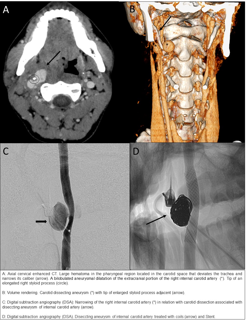
25-year-old previously healthy male who was involved in a motor vehicle accident six months ago with posttraumatic cervicalgia. Now presenting with syncope at Emergency Room. The patient was conscious with odynophagia and neck pain. Exploration by ENT showed a hematoma in the right anterior pillar of the pharynx that displaces the uvula. Steroids were administered and he was admitted at the ENT Department and reexplored 6 hours later. At nasofibroscopy a large hematoma in the posterior wall of the pharynx was observed and emergent cervical CT was requested. Cervical enhanced CT shows a large hematoma in the pharyngeal region located in the carotid space. It deviates the trachea and narrows its caliber. A bilobulated aneurysmal dilatation of the extracranial portion of the right internal carotid artery with dissection is seen. The tip of an elongated styloid process is adjacent to the aneurysm. Findings are in keeping with Eagle syndrome with dissecting aneurysm of the internal carotid artery (stylocarotid syndrome) complicated with rupture and hematoma. The interventional neuroradiologist confirmed these findings with digital subtraction angiography and treated the aneurysm by coil-embolization and a covered stent with total closure. Control by angiography and cervical CT two months later was recommended demostrating resolution of the mass effect and no signs of recanalization, followed by a cervicotomy for resection of styloid processes.
Discusión
Eagle syndrome was described as an elongated styloid process ( greater than 3 cm) or calcified stylohyoid ligament associated with symptoms like neck pain, otalgia or dysphagia. The styloid process is a bony prominence located on the inferior aspect of the temporal bone that extends anteromedially. Stylocarotid syndrome is a low frequency vascular variant from the Eagle syndrome and results from the styloid process compressing the carotid artery. A rare complication of this condition is carotid artery dissection. Carotid artery dissection is caused by disruption of the vessel wall layers with a tear in the intima or rupture of the vasa vasorum with a intramural hematoma, resulting in stenosis or oclusion. If the dissection extends toward the adventitia, it may form a dissecting aneurysm, that becomes a nidus for distal thromboembolism or causes mass effect on adjacent structures. One of the potential life threatening complications is the rupture of the aneurysm.
Conclusión
In patients presenting with carotid dissection it is paramount to think of an elongated styloid process as a potential cause.
Bibliografía
- Rodallec MH, Marteau V, Gerber S, Desmottes L, Zins M. Craniocervical Arterial Dissection: Spectrum of Imaging Findings and Differential Diagnosis. RadioGraphics 2008, 28: 1711-28. - Smoot TW, Taha A, Tarlov N, Riebe B. Eagle syndrome: A case report o