Hospital: Hospital Regional Universitario.
Nº: C2019-733
Aut@r o Autores: L. Sánchez Linares, C. Simón Bejarano, L. Tenorio Tornero.
Presentación
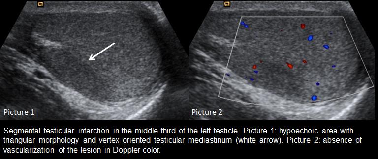
21 year-old male, no medical history of interest went to emergency room with acute left testicular pain, 4 hours of evolution. Does not concern prior traumatic history. At physical examination the left testicle is quietly ascended, without increasing size or changes in coloration, with selective tenderness of the upper pole of the testicle. Requests emergency ultrasound for suspected testicular torsion. In testicular ultrasound was noted an hypoechoic well defined area at the level of the middle portion of the left testicle, with triangular morphology and with vertex directed toward the mediastinum, without mass. The area didn’t showed vascularization at the Doppler color study. The rest of the testicle, as well as the contralateral present normal vascularization. Both epididymides were normal. Hydrocele is not observed. After the findings was performed surgical exploration, where it confirmed the presence of an area at the level of the middle part of the left testicle compatible with segmental testicular infarction. Spermatic cord did not show signs of torque and the rest of the left testicle and the right testicle had a proper coloring. An orchidopexy and subsequent follow up performed by urology and radiology.
Discusión
Segmental testicular infarction is an uncommon pathology, and is presenting clinically as acute scrotum. Sonography shown an hypoechoic area of well-defined margins, triangular morphology, with a vertex oriented testicular mediastinum and absence of vascularization in Doppler color, most often located in the upper portion and half of the affected testicle. These features are highly suggestive of the diagnosis, however, if you have a rounded appearance can simulate a little vascularized tumor, by contrast-enhanced ultrasound can be useful. Main differential diagnoses include: primary testicular tumor, metastases secondary to a process lymphoproliferative or abscess.
Conclusión
Segmental testicular infarction is an uncommon cause of acute scrotum who a radiologist may know recognize to avoid invasive and unnecessary additional tests, as well as the main differential diagnoses
Bibliografía
- D Sommers, Winter T. Ultrasonography evaluation of scrotal masses. Radiol. Clin. North am. 2014, 52 (6): 1265-81. - EA, Khati NJ, Hill MC Akin. Ultrasound of the scrotum. Ultrasound q. 2005, 20 (4): 181-200.