Hospital: Hospital Universitario Virgen Macarena.
Nº: C2019-684
Aut@r o Autores: I. Avilés Vázquez, X.M. Cortés Sañudo, R.A. Domínguez Rodríguez, R.S. Estelles López, P. García Rodríguez, M. Roquette Mateos.
Presentación
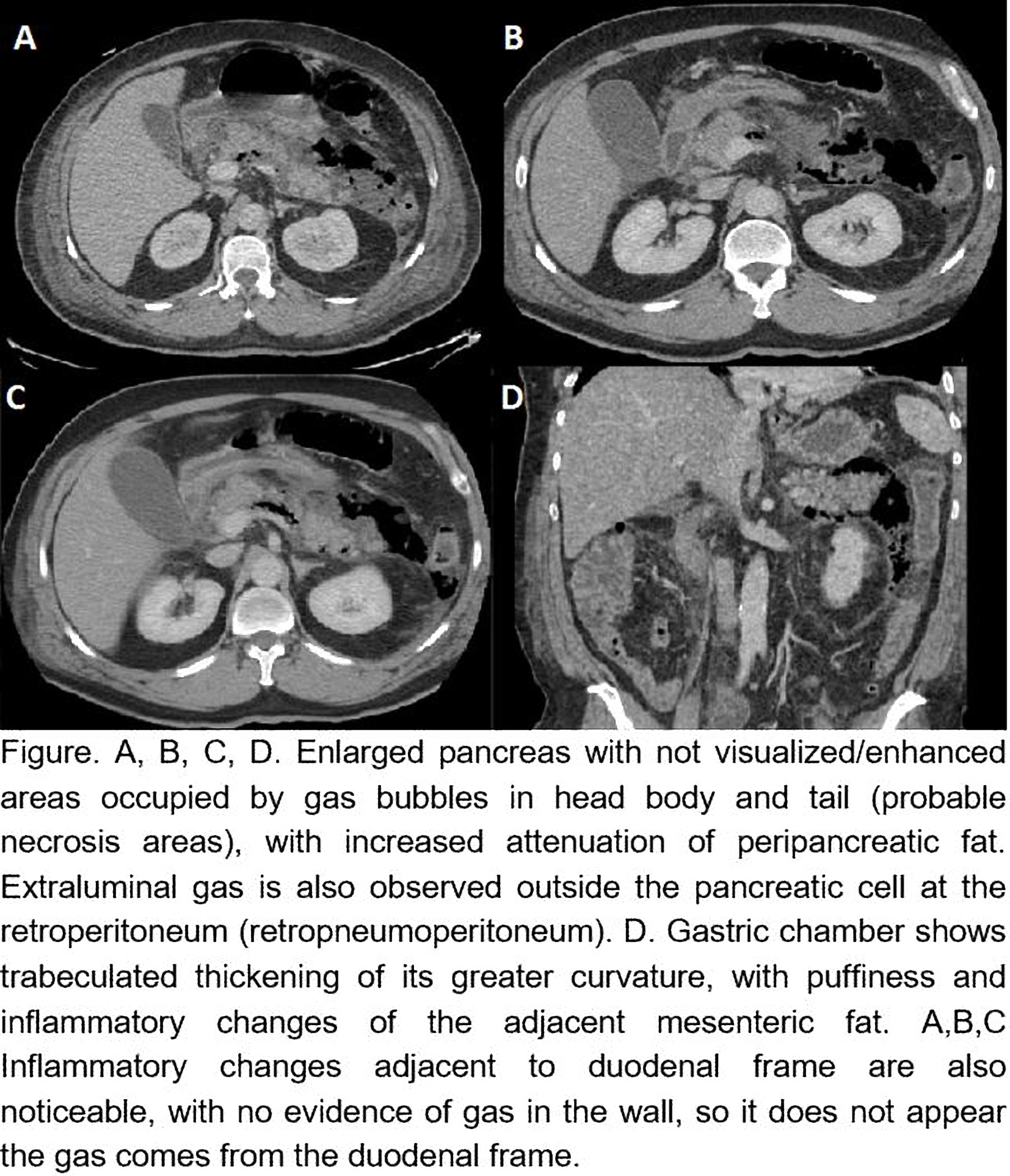
A 58-year-old man consulted for clinical symptoms of severe epigastric abdominal pain accompanied by nausea and bilious vomiting. In the analytical study, significant leukocytosis and hyperamylasemia is highlighted. Abdomen CT with IV contrast, without oral contrast is performed, in which we observe enlarged pancreas with not visualized/enhanced areas occupied by gas bubbles in the head body and tail (probable necrosis areas), with increased attenuation of peripancreatic fat. Extraluminal gas is also observed outside the pancreatic cell at the retroperitoneum (retropneumoperitoneum). Likewise, the gastric chamber shows trabeculated thickening of its greater curvature, with puffiness and inflammatory changes of the adjacent mesenteric fat. Inflammatory changes adjacent to duodenal frame are also noticeable, with no evidence of gas in the wall, so it does not appear the gas comes from the duodenal frame.
Discusión
Acute pancreatitis (AP) is a high mortality rate disease. The revised Atlanta classification distinguishes two morphological types of AP, edematous interstitial pancreatitis and necrotizing pancreatitis, the latter being the worst prognosis. To perform a PA diagnosis, at least the presence of one of the following findings is required: edematous pancreas parenchyma, striation of the peripancreatic fat and/or liquid collection, parenchyma and/or peripancreatic necrosis. Emphysematous pancreatitis is a potentially fatal variant of severe necrotizing AP, defined by the presence of gas in the pancreatic cell and indicates the existence of a bacterial infection, with variable affectation of other tissues by neighborhood and at distance. The detection of retropneumoperitoneum is the clue in the diagnosis of emphysematous pancreatitis. Although the presence of gas in the pancreas is mainly related to infection by gas-forming microorganisms, mainly E. coli, it can also be associated with the formation of an entero-pancreatic fistula, for example, after endoscopic instrumentation or sphincterotomy. The prognosis of this entity is fatal and early radiological detection can influence survival.
Conclusión
Emphysematous AP is a rare and threatening condition for patient's life. The diagnosis of this entity is radiological, based on demonstration of gas in the retroperitoneum in a patient with clinical evidence of pancreatitis. CT is the most sensitive and specific diagnostic method. Although the identification of isolated gas bubbles is not specific to an infectious process and therefore to emphysematous pancreatitis, its presence in anarea of pancreatic necrosis on CT is considered a positive indicator of gas-forming microorganisms presence.
Bibliografía
- Balani A, Dey A, Sarjare S, Chatur C. Emphysematous pancreatitis: classic findings. BMJ Case Reports. 2016,:bcr2016217445. - Grayson D, Abbott R, Levy A, Sherman P. Emphysematous Infections of the Abdomen and Pelvis: A Pictorial Review. RadioGraphics.