Hospital: Complejo Hospitalario de Navarra.
Nº: C2019-110
Aut@r o Autores: N. Alberdi Aldasoro, P. Lopez Sala, C. Saavedra Gutierrez, N. Alonso Ordas, G. Unzue Garcia-Falces, L. De Llano Ibisate.
Presentación
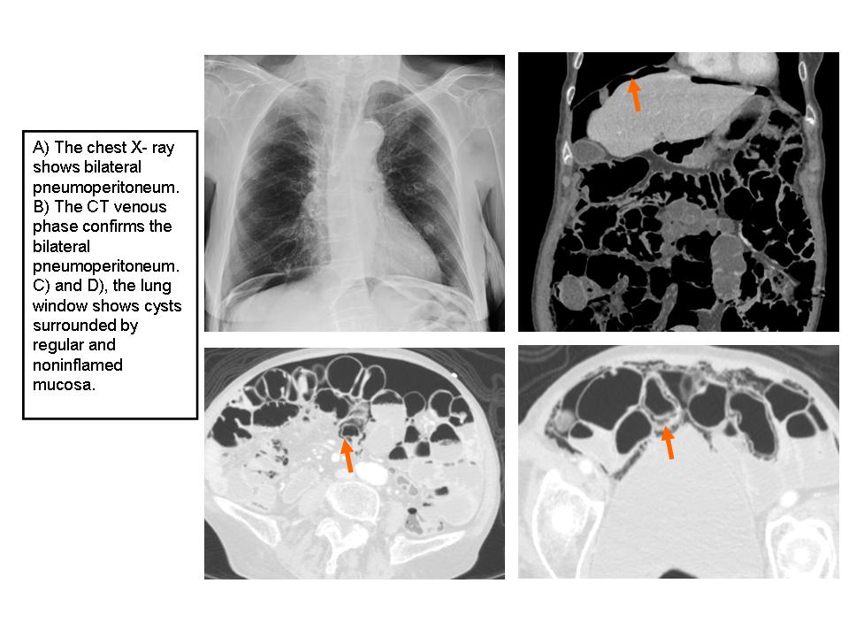
90 years old woman comes to the emergency room with dizziness, asthenia and general discomfort. The chest x-ray shows bilateral pneumoperitoneum but the patient denies acute abdominal pain. On examination, her abdomen was distended but soft, with normal bowel sounds. The abdomen CT shows pneumoperitoneum and parietal pneumatosis in small bowels.
Discusión
Pneumatosis cystoides intestinalis (PCI) is a rare condition characterized by the accumulation of submucosal or subserosal gas-filled lesions in the gastrointestinal tract. Although the cause of PCI appears to be multifactorial, the exact cause is not known but various coexisting diseases, including respiratory diseases, inflammatory bowel diseases, collagen diseases, malignancies, and infectious diseases, may be associated with the onset of PCI.They are most often located in the small bowel, less frequently in the large, and quite rarely in the stomach or omentum (1). Patients may be asymptomatic or complain of pain and abdominal distension, diarrhea and rectal blood loss. Most often affects males between the fifth and sixth decades of life. CT imaging is the gold standard procedure for the diagnosis. The CT image shows air in multiple small cysts within the wall of the colon and sometimes subserous bubbles can break spontaneously with pneumoperitoneum. The main differential diagnosis is with intestinal ischemia where we can see bowel wall thickening, altered contrast mucosal enhancement, dilated bowel, soft tissue stranding, ascites, and the presence of portal air. Although treatment will be varied according to the severity and cause of the ischemia, in general treatment is surgical. In contrast the PCI does not require any specific treatment, so It is important to distinguish them. Therefore, the correlation with clinical history, physical examination, and laboratory test results are the best indicators for diagnosis (2).
Conclusión
The clinical presentation of PCI is variable and most patients are asymptomatic, so the radiologist must know this entity and considers the diagnosis in the presence of pneumoperitoneum in order to correctly manage of the patients. 6.
Bibliografía
- Ho LM, Paulson EK, Thompson WM. Pneumatosis intestinalis in the adult: benign to life-threatening causes. AJR Am J Roentgenol. 2007,188:1604–1613. - LL W, Yang YS, Dou Y, Liu QS. A systematic analysis of pneumatosis cystoids intestinalis. World J Gast