Hospital: Hospital Universitario Ramón y Cajal.
Nº: C2019-541
Aut@r o Autores: I. Pecharromán De Las Heras, C. Sempere Ortega, A. Vicente Bártulos, C. Campos Ferrer, C. Picón Serrano, E. García Casado.
Presentación
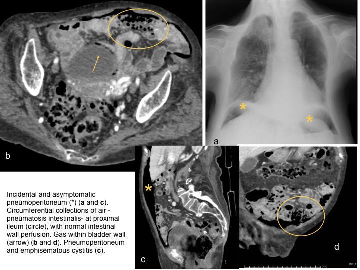
A woman aged 93 gets a chest radiograph to rule out pulmonary infection. Incidental asymptomatic pneumoperitoneum is found, and a contrast-enhanced abdominopelvic CT is recommended. At CT we found pneumoperitoneum and extraperitoneal gas, little ascites, bubbles of air within a segment of ileum wall (with adequate enhancing pattern), and gas within bladder wall (no clinical signs of cystitis). There are jejunum and colonic diverticula (not shown), gas at uterine cavity and good opacification of mesenteric vessels.
Discusión
This is a case of idiopathic pneumatosis cystoides intestinalis or pneumatosis intestinalis (PI) associated with both asymptomatic pneumoperitoneum and emphysematous cystitis. The patient was hospitalized to be treated with oxygen therapy and intravenous antibiotics (for respiratory infection), with good outcome. Pneumatosis intestinalis can be an incidental finding associated with a benign etiology, whereas in others it implies a life-threatening condition. Incidence is uncertain since asymptomatic patients never come to clinical attention. It is idiopathic up to 15%. Pathogenesis is poorly understood, with three main hypotheses considered: the mechanical (gas dissects into the wall of the bowel from either the luminal or through the serosal surfaces), the bacterial (gasforming bacteria gaining access to the submucosa through breaches in the mucosa) and the biochemical theory (excessive production of hydrogen gas through fermentation of carbohydrates and other food). None of them are conclusive [1]. Pneumoperitoneum is probably secondary to the rupture of cysts, but it does not imply clinically worrisome PI.Emphysematous cystitis is uncommon, and often incidental, more frequent in females with diabetes mellitus [2]. It can be associated with noninfectious sources of air such us pneumatosis cystoides intestinalis [3]. A contrast-enhanced abdominal CT must be done when PI or pneumoperitoneum are diagnosed at abdominal radiograph, to establish the diagnosis, determine the underlying etiology, and diagnose complications. Findings include collections of air in the bowel wall. Decreased mural contrast enhancement and associated portal venous gas are suggestive of intestinal ischemia, although 30% of this patients have a benign idiopathic cause. Management includes emergent surgical exploration (if clinical and metabolic signs of ischemia are present) and medical management (antibiotics, oxygen therapy and diet). Asymptomatic patients do not require any additional therapy.
Conclusión
Most patients with PI are asymptomatic. Complications, such as pneumoperitoneum or emphysematous cystitis, may be asymptomatic too. It is mandatory to rule out life-threatening conditions.
Bibliografía
- Dinkel HP, Lourens S, Brehmer U, Pfammatter R, Triller J, Vock P. Emphysematous cystitis in a patient presenting with paradoxical arterial embolism and intestinal mycobacteriosis without evidence of diabetes. Eur Radiol 2001,11(2):246-9. - Goldberg E.