Hospital: Hospital Universitario San Cecilio. ·
Nº: C2019-259
Aut@r o Autores: L. Guirado Isla, C. Dávila Arias, J. Miras Ventura, I. Garrido Márquez, F. Briones Bajaña, M. Fernández Conesa.
Presentación
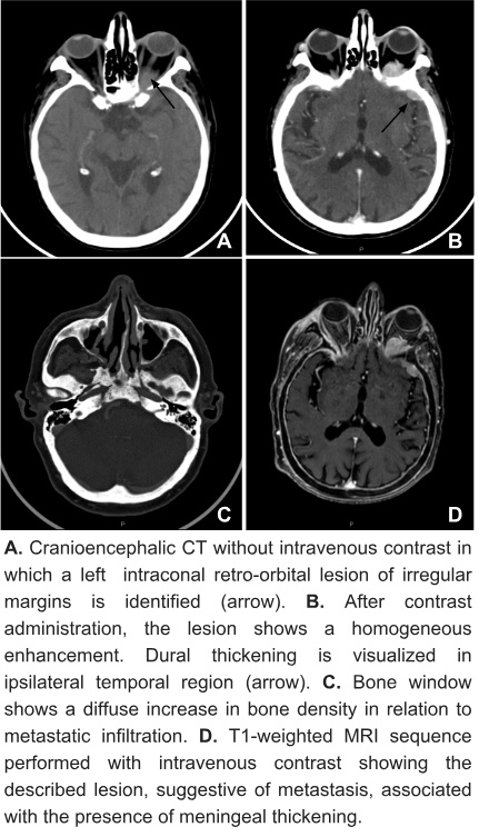
78-year-old male with antecedent of metastatic prostate cancer presented with history of loss of vision, redness and proptosis in the left eye associated with episodes of fluctuating diplopia. He described sudden onset of the clinic five days ago and its progressive increase. Clinical examination showed proptosis, medium mydriasis, slight conjunctival redness and immobility of the left eye with preserved visual acuity. Pre- and postcontrast cranioencephalic CT was performed in which an intraconal lesion with soft tissue density was identified. It seemed to follow the axis of the optic nerve and contacted the oculomotor muscles determining ipsilateral proptosis. In addition, there was a diffuse increase in cranial bone density. After the administration of intravenous contrast, a homogeneous enhancement of the described lesion was observed, as well as its continuity with an intracranial dural thickening of nodular morphology. The study was subsequently completed with cranial MRI confirming the diagnosis of retro orbital metastasis of prostate adenocarcinoma with diffuse metastatic bone involvement.·
Discusión
Orbital neoplasms are often primary or result from the invasion of tumors located in proximity. Nevertheless, metastatic lesions constitute between 1% and 13% of them, being the most frequent those of breast cancer followed by prostate carcinoma, melanoma and lung cancer. Symptoms usually show a rapid evolution (from weeks to months) and habitually consist of proptosis, alterations of ocular motility, pain, diplopia or decreased vision. It may be the first manifestation of a neoplasic process and the prognosis is generally poor. CT and MRI are fundamental in the assessment of this type of pathology, although CT shows higher sensitivity to detect associated bone affectation and MRI is more useful discriminating between tumor and inflammatory nature. The most common radiological finding is an unilateral intraorbital mass that can be accompanied by bone, muscle and fat or only muscle involvement, which can lead to several types of primary neoplasia (prostate cancer, breast cancer or melanoma respectively). Differential diagnosis with non-neoplastic infiltrative pathologies may be a challenge in imaging tests: in particular, with respect to conal metastases both thyroid ophthalmopathy and orbital pseudotumor should be considered.
Conclusión
Orbital metastatic lesions are a posible diagnosis to be taken into account in patients with compatible symptoms, especially in those with known oncological disease. Differentiating between tumor or inflammatory origin can be complex in imaging tests, of which CT and MRI are considered the techniques of choice.·
Bibliografía
- Tailor TD, Gupta D, Dalley RW, Keene CD, Anzai Y. Orbital neoplasms in adults: clinical, radiologic and pathologic review. Radiographics 2013, 33 (6): 1739-1758. - González F, López C. Metástasis orbitarias: Serie de cuatro casos y