Hospital: Hospital de la Santa Creu i Sant Pau.
Nº: C2019-47
Aut@r o Autores: C. Sitges, N. Martínez, B. Tintaya, J. Palmer.
Presentación
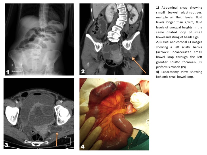
A 49-year-old female was admitted to the hospital presenting continuous lower abdominal pain, nausea and vomiting. The patient had unremarkable past surgical history, with no prior abdominal surgery, and no relevant past medical history. On examination, positive findings included marked abdominal distension, generalized abdominal tenderness and constipation. Important negative findings included no hernia and no signs of peritonism. Upright abdominal radiograph was performed showing signs of small bowel obstruction. To identify the cause of the small bowel obstruction, a CT scan of the abdomen with intravenous contrast was performed. The images revealed small bowel obstruction secondary to incarcerated sciatic hernia. Surgical exploration revealed the sciatic hernial sac containing ischemic small bowel loop and atrophic left ovary. Resection of the ovary and small bowel with hernioplasty were performed.
Discusión
Sciatic hernia defined as protrusion of the peritoneal sac and its contents through the greater or lesser sciatic foramen. It is a very rare entity (<0,01%), but its incidence is significantly growing in the past years due to the widespread use of radiological investigations and laparoscopic techniques. It is more prevalent in females and its etiology is unclear. Probably a genetic predisposition, an increased space between piriformis muscle and sacrospinous ligaments and a chronic raised intraabdominal pressure could be considered two of the most important risk factors. Suprapiriformis hernia is said to be the most common type, which corresponds with the findings of the present study. The clinical presentation is various, it may present with obscure pelvic pain, intestinal obstruction, life-threatening gluteal sepsis, or as an asymptomatic, reducible mass that distorts the gluteal fold. Small sciatic hernia can remain hidden behind the gluteus maximus muscle. The diagnosis requires imaging studies in such cases. Ultrasonography and CT are the imaging modalities commonly used to diagnose sciatic hernia, although MRI can be used in cases in which entrapment of the sciatic nerve is suspected.Treatment of sciatic hernia is always surgical and requires prosthetic reinforcement for a best result.
Conclusión
In conclusion, the diagnosis of an incarcerated sciatic hernia can be difficult, but awareness of this unusual entity in patients presenting small bowel obstruction and performing imaging studies make it possible.
Bibliografía
- Colombo, F., Calcagno, P., Crespi, M., Bonzanini, O., Sampietro, G. M., & Foschi, D. Laparoscopic Repair of a Sciatic Hernia Containing the Ipsilateral Ovary: Case Report and Review of the Literature. Journal of Laparoendoscopic & Advanced Surgical Tech