Hospital: Hospital Universitario San Cecilio.
Nº: C2019-381
Aut@r o Autores: C. Martínez Martínez, I. Garrido Márquez, A. Milena Muñoz, J. Miras Ventura, L. Díaz Rubia, L. Guirado Isla.
Presentación
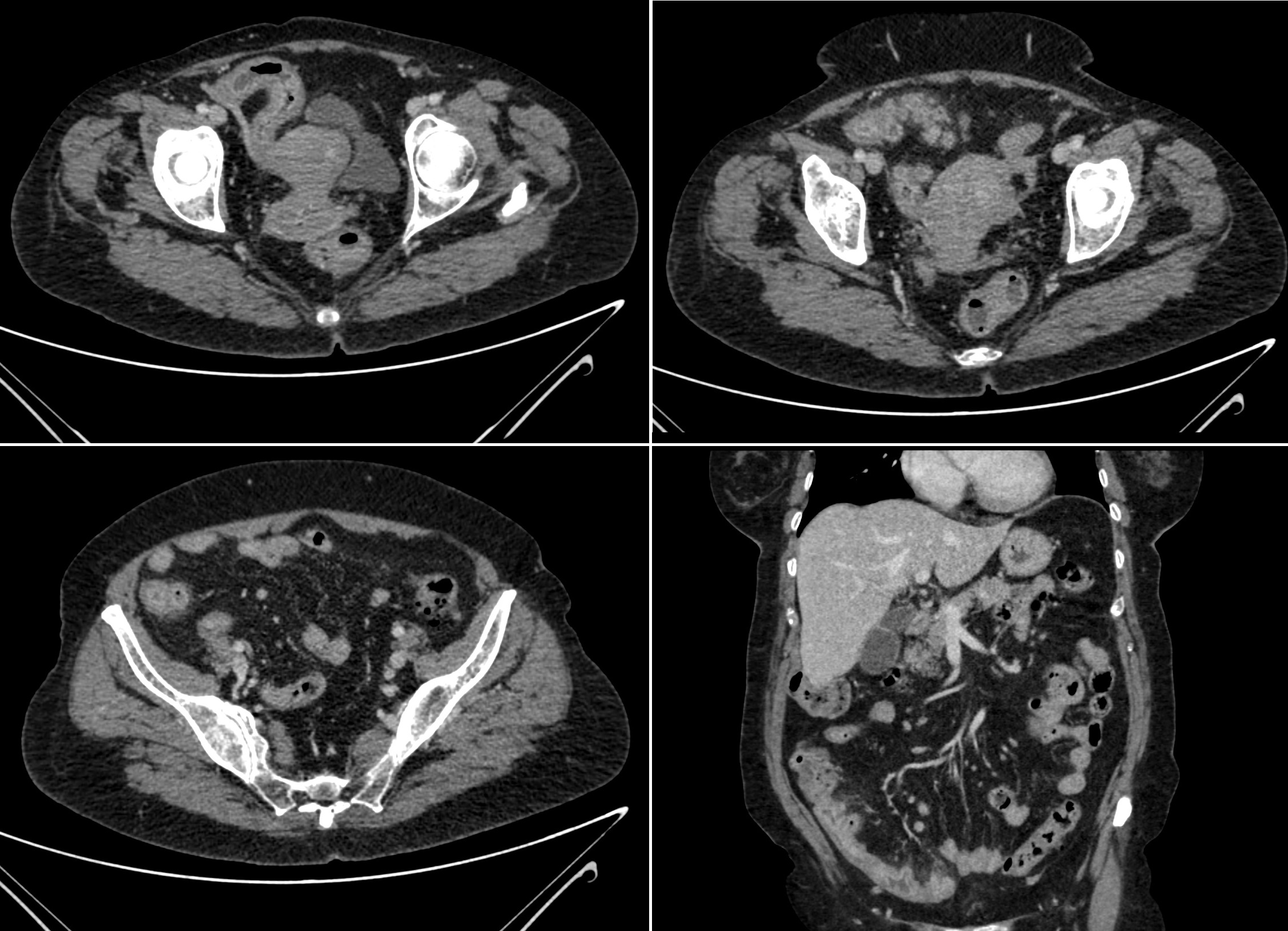
A 63 year-old patient with colonic diverticulosis came to the Emergency Department for acute abdominal pain. Blumberg and Rovsing’s signs were positive and Murphy and kidney percussion were negative. The main findings in the laboratory test were PCR 309, leukocytes 6140, neutrophils 75.5%, total bilirubin 1.36 with direct bilirubin 0.51 and indirect 0.85. Portal venous-phase contrast-enhanced abdominal CT was performed and diverticular images in distal ileum with parietal thickening and mucosal enhancement, stranding of adjacent fat and reactive lymphadenopathies were seen, findings very suggestive of ileum diverticulitis.
Discusión
Ileum diverticulitis is a rare pathology. The most frequent diverticula are those located in colon, followed by duodenum, esophagus, stomach, jejunum and ileum. They are usually asymptomatic but there may be complications such as diverticulitis, perforation, obstruction, abscess, anemia or volvulus. When ileum diverticulitis affects distal ileum, differential diagnosis must be made with acute appendicitis. Laboratory tests are also nonspecific, being the leukocytosis the most common finding followed by elevated CRP. Portal venous phase contrast-enhanced CT is preferred and it is not necessary to administer oral contrast, not being recommended in patients with suspected acute diverticulitis. The findings of an ileum diverticulitis are very similar to colonic diverticulitis: parietal thickening associated with inflammatory changes such as stranding of the adjacent fat and presence of free fluid among others.
Conclusión
The ileum diverticulitis is a quite infrequent pathology and scarcely described in the scientific literature. Symptoms and laboratory tests are very nonspecific and can simulate acute appendicitis. Portal venous phase contrast-enhanced CT is the preferred imaging modality and the findings are very similar to colonic diverticulitis.
Bibliografía
- Transue DL, Hanna TN, Shekhani H, Rohatgi S, Khosa F, Johnson JO. Small bowel diverticulitis: an imaging review of an uncommon entity. Emerg Radiol. 2017 Apr,24(2):195-205. - Jeong J, Hong SS, Hwang J, Kim HJ, Chang YW. Acute diverticulitis of the termi