Hospital: Hospital Universitario de Móstoles.
Nº: C2019-147
Aut@r o Autores: C. Romero-Martínez, E. Antón Pascual, C. Diego Hernández, J. Herrador Benito.
Presentación
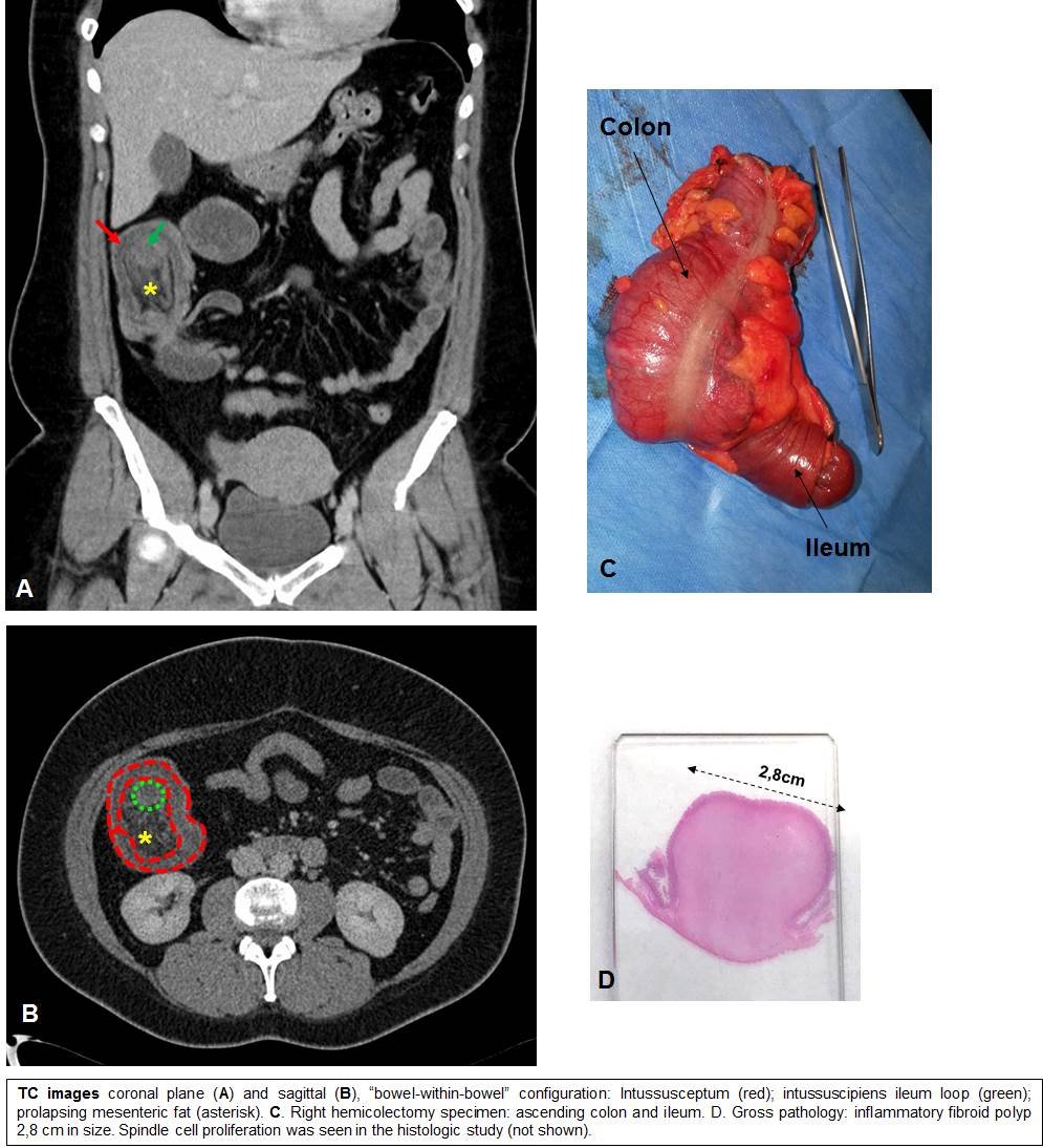
A 37 years old woman presented to the Emergency Department with colicky abdominal pain for two hours, focused on the right upper quadrant. She related nausea and alimentary vomits, denied diarrhoea and temperature. The patient’s surgical history included an appendicectomy and a C-section, with no other relevant medical history. She had also undergone an MR- Cholangiography exploration 6 months before this episode (for diffuse postprandial abdominal pain), which was diagnosed with a probable polyp in the main biliary duct. Laboratory tests revealed a high white blood cell count with neutrophilia, CRP was normal and no other laboratory abnormalities were detected. Abdominal X-ray showed no alterations. The CT scan showed a regular mural thickening of the ascending colon and caecum with a long intussucepting segment of the terminal ileum. Vessels and mesenteric fat could be seen into the intussusception producing a ` bowel-within-bowel ´ pattern. The patient was diagnosed with ileocecal intussusception with no apparent lead point.
Discusión
The woman underwent an emergency laparotomy which proved the intussuscepting ileum through the incompetent ileocaecal valve. A right hemicolectomy was performed as a surgical reduction of the intussusception failed. The pathology exam showed a polyp-like submucosal based lesion 2,8 x 2,5 cm in size, with a spindle cell proliferation in an oedematous stroma with inflammatory cells (lymphocytes, plasma cells particularly and eosinophils). These findings are consistent with inflammatory fibroid polyp in the terminal ileum. The mucosa adjacent to the tumour presented ischemic changes with erosions and ulcerations related to the intussusception. The patient had a satisfactory recovery and was discharged a week after the surgery. Key Learning Points CT imaging is the best technique to diagnose bowel invagination, especially in the Emergency room. Radiological findings of intussusception may be non-specific, although typical signs can be seen such as the “bowel-within-bowel” configuration.
Conclusión
Intussusception in adults is a rare condition, and an underlying lesion must be investigated since, in adults, the lead point is usually a malignant neoplasm. Inflammatory fibroid polyps are uncommon benign lesions of the gastrointestinal tract which usually are indistinguishable from other bowel masses and, as other intraluminal lesions, cancause obstruction and intussusception.
Bibliografía
- Choi SH, Han JK, Kim SH, Lee JM, Lee KH, Kim YJ, et al. Intussusception in adults: From stomach to rectum. Am J Roentgenol. 2004,183(3):691–8. - Harned RK, Buck JL, Shekitka KM. Inflammatory fibroid polyps of the gastrointestinal tract: radiologic eval