Hospital: Hospital Universitario Puerta Hierro.
Nº: C2019-468
Aut@r o Autores: M. Tuñón Gómez, R. Ruiz Peralbo, M. Ibnoulkhatib, M. Collado Torres, Y. Garcia Hidalgo, B. Brea Álvarez.
Presentación
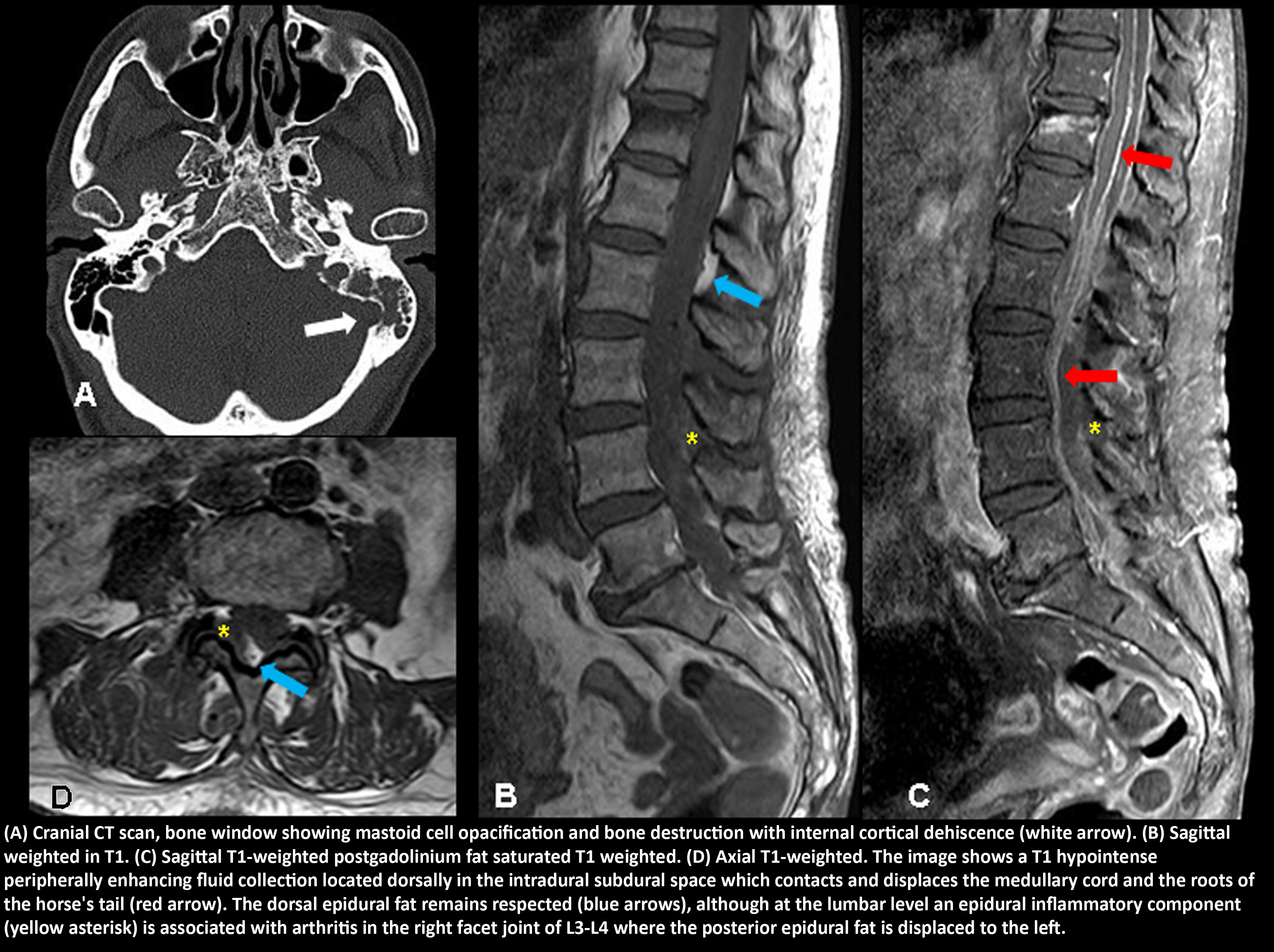
The patient is a 74-year-old woman with arterial hypertension and type 2 DM. She was brought to our emergency unit with a low level of consciousness. According to her family, she experienced a discomfort in her left ear during the last two months. It progressively increased and caused cervical pain. The patient was admitted to the ICU. Cranial CT showed a coalescing otomastoiditis. We suspected an otogenic meningitis. A difficult lumbar puncture was performed. Due to the possibility of spinal abscess or hematoma, an urgent brain MRI was performed (not shown) showing meningitis and purulent ventriculitis. An urgent MRI of the entire spinal column was also performed and showed a subdural dorsal empyema, lumbar facet arthritis, myositis and intramuscular collections in cervical and lumbar paravertebral musculature (not shown). Streptococcus pneumoniae was isolated in otic aspiration and in CSF. The patient was treated with myringotomy, later mastoidectomy, ventricular drainage and intrathecal and intravenous antibiotic therapy with a good improvement. The patient recovered an independence in her daily life.
Discusión
Acute otitis media is common in children. In the adult population it represents 1% and occurs during the ages between 35 and 44 years. It is mostly caused by Streptococcus pneumoniae. Complications in adults and diabetes mellitus as in our case is a predisposing factor. The extratemporal manifestations are referred as neuroinvasive diseases, the mechanism of dissemination may involve the dehiscence of the bone tegmen of the roof of the middle ear cavity. When MRI is requested, the suspicion is lumbar epidural abscess / hematoma due to the difficulty of the puncture. The MRI findings revealed a greater severity of the process than expected, neuroinvasive evidence of otitis, meningitis, ventriculitis, and subdural empyemas, as well as muscular and facet joint inflammatory signs. Spinal subdural abscess is a very rare entity.
Conclusión
Neuroimaging is necessary to evaluate extra-temporal complications, especially in cases of severe neurological symptoms and decreased consciousness. It requires a rapid recognition of the complications of AOM and an early participation of a multidisciplinary care team. The observation of the location of the posterior epidural fat is a key finding to determine the presence of spinal subdural empyemas.
Bibliografía
- Patel KM, Johnson JE, Boxerman JL, Nau GJ. Group A streptococcus acute otitis media progressing to neuroinvasive disease in adults. IDCases. 2018,12: 161-164. - Velissaris D, Aretha D, Fligou F, Filos KS. Spinal Subdural Staphylococcus Aureus Abscess: