Hospital: Hospital Universitario Virgen Macarena.
Nº: C2019-686
Aut@r o Autores: X. Cortés Sañudo, R. Estelles López, E. Rangel Villalobos, C. Pérez Ramírez, I. Avilés Vázquez, T. Busquier Cerdán.
Presentación
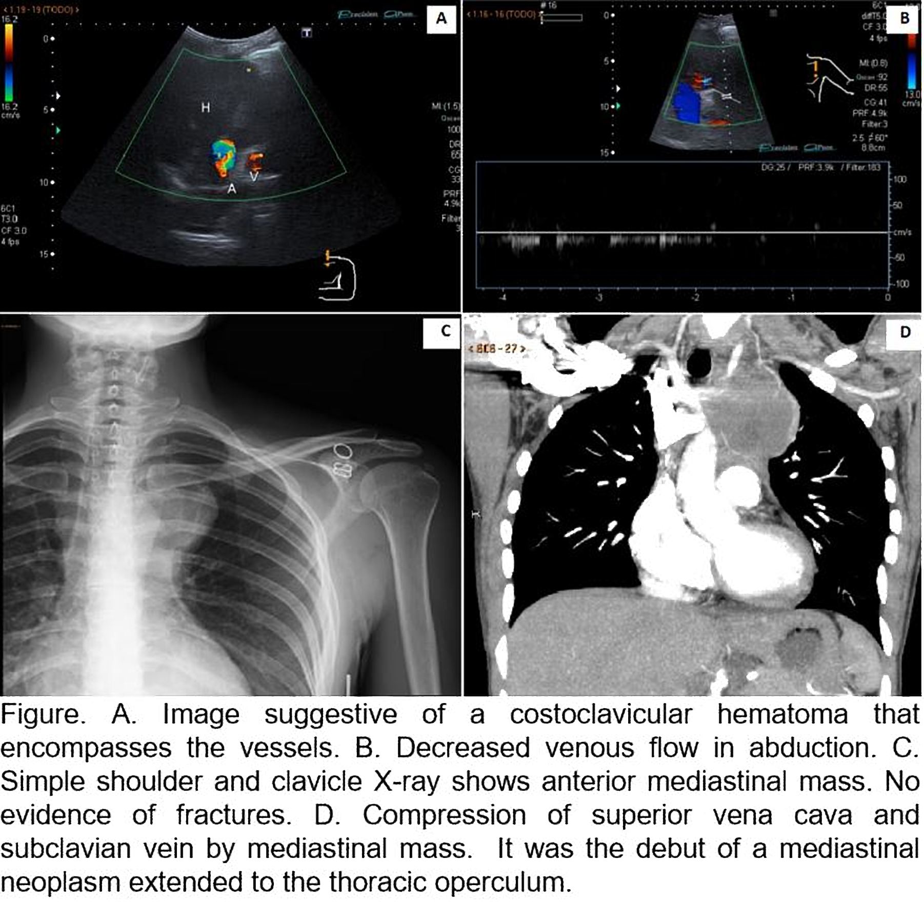
A 37-year-old woman with a clinical history of accidental trauma over left suprasternal area and secondary upper left extremity (ULE) edema: In Doppler ultrasound (DUS) we found a yugulo-subclavian DVT and on left costoclavicular space, a suggestive image of large soft tissue hematoma. On the dynamic ultrasonographic study at hyperabduction, we found a clear decrease in venous caliber and decreased venous flow in abduction, compatible with key findings on Thoracic Outlet Syndrome (TOS). In the complementary study, chest X-ray and CT angiography, showed an anterior mediastinum mass making extrinsic compression on the superior vena cava (SVC) and a secondary left common carotid artery sharpening. After the immunohistochemistry study, it was shown to correspond with a follicular dendritic cell sarcoma (FDCS).
Discusión
FDCS are extremely rare lymphoid neoplasms. They primarily affect lymph nodes with occasional extranodal involvement. The definitive diagnosis requires immunohistochemistry. Its clinical behavior, its treatment, as well as its evolution are little known. The interest of our case relies on its debut after a banal traumatism and the associated symptomatology, a TOS with venous involvement by a yugulo-subclavian DVT. It should It be remembered that despite the great DUS diagnostic power, this technique does not allow the entire evaluation of the thoracic operculum, specially, the pulmonary apex analysis, so it is essential to complement the ultrasound diagnosis with a CT angiography to make an overall thoracic operculum study. The most relevant findings TOS are Vps duplication in the subclavian artery and loss of respiratory dynamics in the subclavian vein at 90° abduction due to narrowing of the vessel. Abduction beyond this angle leads to a decrease in flow, that is, a decrease in Vps in the subclavian artery due to a pre-occlusion phase. At hyperabduction, the absence of flow indicates total occlusion. This is observed in severe cases.
Conclusión
DUS dynamic imaging provides an important first-level diagnostic tool for the radiologist in case of a suspected diagnosis of TOS. This image technique can provide valuable and accurate information about the existence and degree of stenosis at differentvascular levels. There is a group of uncommon tumor pathology that can lead to a TOS by compression, where isolated cases of desmoid tumor, lipoma or hemangioma of the first rib have been described, among others.
Bibliografía
- Raptis C, Sridhar S, Thompson R, Fowler K, Bhalla S. Imaging of the Patient with Thoracic Outlet Syndrome. RadioGraphics. 2016,36(4):984-1000. - Eliahou R, Sosna J, Bloom A. Between a Rock and a Hard Place: Clinical and Imaging Features of Vascular Com