Hospital: Radiologist Hospital Alto Guadalquivir, Chief of Radiology Department.Hospital Alto Guadalquivir.
Nº: C2019-220
Aut@r o Autores: A. Gampel Cohen, A. Palma Baro, V. Palomo Gallego, A. Higuera Higuera, I. Rivera Salas, M. Jaén Reyes.
Presentación
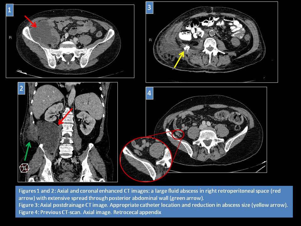
A 65-year-old male presented to Emergency Department (ED) with thoracolumbar painful mass from some days before. Patient reported a medical history of jejunal adenocarcinoma and peritoneal carcinomatosis on treatment with chemotherapy. Physical examination revealed a lumbar mass which was indurated, hot and attached to deep planes. Laboratory tests showed leukocytosis.Soft-tissue ultrasound was requested and showed thickness of subcutaneous fatty tissue with diffuse hyperechogenicity in a very deep extension. Contrast enhanced CT-scan was performed and identified in right retroperitoneal space a large fluid collection with ring enhancement consistent with abscess. Collection had mass effect on the right psoas muscle and extensive spread through posterior abdominal wall. Also it was in contact with ascending colon and cecum but without thickening of bowel wall, pneumatosis or fat stranding. Appendix was not identified so the origin of infection was uncertain. Prior studies from the patient were reviewed and showed that appendix was in retrocecal location and it might be the cause of the disease. CT-guided percutaneous drainage was performed and purulent fluid was removed with positive Proteus mirabilis culture. Postprocedure CT-scan demonstrated an appropriate catheter location and reduction in abscess size. Patient was deemed unfit for surgery because of comorbid disease and favourable clinical evolution. However, our patient died two months later secondary to another complication of his malignant disease.
Discusión
Immunocompromised patients include those undergoing medical treatment for a malignancy. These patients may visit ED for multiples causes like treatment toxicity or opportunistic infections. However, we must not forget that these patients may develop other pathologies that, due to their immunodeficiency state, can course in a different way in symptoms (atypical pain, atypical location…) or evolution. In the same way, physical examination may be confused so imaging techniques play a fundamental role to establish a correct diagnosis and treatment. If a patient who is receiving chemotherapy for malignant disease reports an abdominal right-side pain, the most common clinical suspicion is neutropenic colitis. In our patient, there were no findings of bowel inflammation in CT-scan so an atypical acuteappendicitis was assumed like the origin of retroperitoneal abscess.Key learning points: atypical appendicitis, abscess, immunosuppressed
Conclusión
Acute appendicitis is one of the most common diagnosis in emergency radiology. Generally it is detected like a right-side lower abdominal pain. However, in immunosuppressed patient it can have different symptons and evolution as in our patient who debuted with a retroperitoneal abscess.
Bibliografía
-Purysko AS, Remer EM, Leao-Filho HM, Bittencourt L, Lima RV, Racy DJ. Beyond appendicitis: common and uncommon gastrointestinal causes of right lower quadrant abdominal pain at multidetector CT. RadioGraphics 2011,31:927-947. - .Viswanathan C, Truong M