Hospital: Hospital Universitario Virgen de las Nieves.
Nº: C2019-191
Aut@r o Autores: M. Rabadán Caravaca, J. Parejo Santaella, M. Pérez García, M. García Roa, G. López Milena.
Presentación
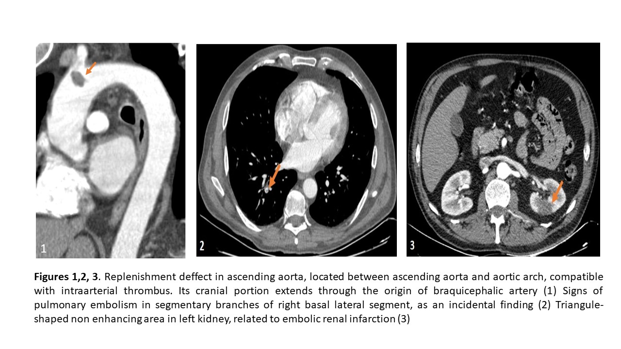
We present a 59 years old man with acute right arm pain not related with previous trauma. A lack of right radial pulse was detected. Our patient didn't have any important personal history or cardiovascular risk factors. A non-contrast toracic CT and CT-Angiography were performed to evaluate acute aortic syndrome, showing the following findings: - Replenishment deffect in ascending aorta, located between ascending aorta and aortic arch, compatible with intraarterial thrombus. Its cranial portion extends through the origin of braquicephalic artery (Figure 1) - Signs of pulmonary embolism in segmentary branches of right basal lateral segment, as an incidental finding (Figure 2) - Triangule-shaped non enhancing area in left kidney, related to embolic renal infarction (Figure 3) The therapeutic management of this patient was anticoagulation, due to the progressive dissapearing of the arm pain. Nevertheless, the thrombus remained in posterior CT control, so a surgical intervention was decided. Before making it, a coronary CT study was made, finding the complete resolution of the thrombus in the 11th day of evolution.
Discusión
Despite most systemic embolisms are detected in patients with left heart disease, aortic thrombus is another important cause of arterial embolism. The risk factors for this entitie are arteriosclerosis, arterial dissection, advanced age, trauma, malignant tumors, and hemostatic disorders. In some cases, an aortic thrombi may be an incidental finding, its natural course is not well known and the pathophysiology is undefined. The most frequent location of thoracic aorta thrombus is the aortic isthmus and the distal portion of aortic arch at the opposite side to the origin of the subclavian artery. The presence of pedunculated thrombi in the aortic arch is not common. According to a retrospective computerised research, in which the radiological findings were correlated with clinical manifestations, the most common radiological finding in CT was a contrast filling defect with narrow base of attachment without calcification, more frequent in symptomatic patients. Surgical removal of the thrombus is followed by good clinical results. Conservative treatment with anticoagulation should be considered in asymptomatic, inoperable or high-risk patients.
Conclusión
Aortic-floating thrombus should be actively sought and excluded when performing CT angiography for emboli. - Surgery remains the best treatment option in symptomatic patients, but it might be neccesary to individualize this decision.
Bibliografía
- Klang E, Kerpek A, Soffer S, Zlotnik M, Shimon O, Rimon U et al. CT imaging features of symptomatic and asymptomatic floating aortic thrombus. Clinical Radiology. 2018,73(3):323.e9-323.e14 - Noh TO, Seo PW. Floating thrombus in aortic arch. Korean J Th