Hospital: H G U R S.
Nº: C2019-45
Aut@r o Autores: M. Ojados Hernández, L. Sánchez Alonso, L. Abenza Oliva, L. Alemañ Romero, A. Puerta Sales, P. Alemán Díaz.
Presentación
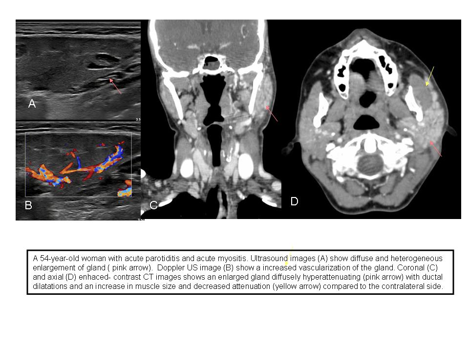
A 54-year-old woman presented with facial tumor, dysphagia and fever of 5 days evolution.In CT of the neck, an enlargement of the left parotid gland is observed with duct dilatations and the Stenon duct. Inflammatory changes in the adjacent soft tissues and signs of myositis in the left masseter.
Discusión
Acute inflammation of the parotid gland is most commonly caused by infections. Acute parotitis tends to manifest with painful facial swelling and tenderness at the angle of the mandible and may be associated with fever and leukocytosis. It can be complicated by an abscess, soft tissue infection. Myositis is a rare pathology since the muscles are organs resistant to infection. The diagnostic keys are: US: The standard of reference for diagnosing acute parotitis. The parotid gland may show diffuse and heterogeneous enlargement with foci of salivary secretions and lymph nodes. At Doppler US, there is increased vascularization of the gland. CT: The role of CT is to confirm complications, usually abscesses. At enhanced CT, the gland appears diffusely hyperattenuating and may show internal fluid collections. Myositis is observed as an increase in muscle size, decreased attenuation, heterogeneous enhancement, and effacement of adjacent fatty plans. MRI: enlarged gland with altered signal intensity, hyperintensity in the T2 and an important enhaced contrast. Myositis is observed as an increase in muscle size, hyperintensity of muscle and the fascia surrounding it in T2.
Conclusión
The standard of reference for diagnosing acute parotitisis is the ultrasound, however CT or MR are necessary if suspect complications. Myositis is an infrequent complication because the muscles are resistant to infection.
Bibliografía
- Inajeros EJ, Navallas M, Tolend M, Suñol M, Rubio-Palau J, Albert A, et al. Imaging evaluation of pediatric parotid gland abnormalities. Radiographics 2018, 38:1552-1575. - Capps EF, Kinsella JJ, Gupta M, Bhatki AM, Opatowsky MJ. Emergency imagin