Hospital: Hospital de la Santa Creu i Sant Pau.
Nº: C2019-223
Aut@r o Autores: A. Corujo, S. Mazzini.
Presentación
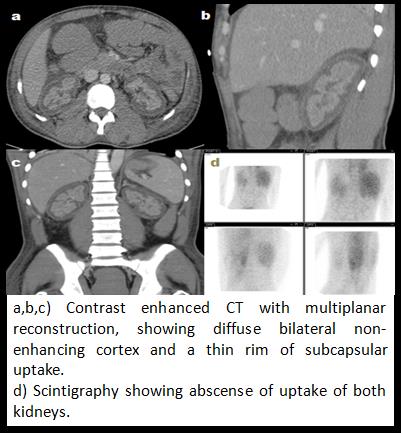
An adult senegalese of unknown age went to the emergency room due to vomiting, pain in the right hypochondrium and oliguria for the past 24 hours. Laboratory findings included acute renal failure and metabolic acidosis. An ultrasound was initially requested, showing kidneys of normal size and cortical thickness with diffuse hyperechogenicity of the cortex (images not available). Due to the lack of improvement of the renal failure with medical treatment, the patient started hemodialysis and an abdominal CT with contrast was performed. The CT showed normal-sized kidneys with bilateral diffuse contrast hypoenhancement in the renal cortex, whit a subcapsular contrast-enhanced "ring", findings compatible with acute cortical necrosis. Toxicological studies and multiple serologies were performed without positive results for any of the tests. A DSMA scintigraphy 3 months later showed abscense of scintigraphic uptake of the kidneys due to severe parenchymal disorder.
Discusión
Renal cortical necrosis is characterized by a patchy or diffuse destruction of all elements of the renal cortex due to a significant decrease in renal arterial perfusion caused by vasospasm and microvascular damage, although the whole picture of its physiopathology is unknown. (1) It is a rare pathology in developed countries (2% of acute renal failure) and is mainly associated with obstetric complications (1). When the pathology is not-obstetric related, the causes are varied: haemolytic uremic syndrome, sepsis, burns, pancreatitis, diarrhea associated with shock, drugs and toxins. (1) When there is diffuse compromise of the cortex, the evolution is towards an irreversible chronic renal failure. (1) Findings in abdominal-CT at a porto-venous phase include a non-enhancing cortex with enhancement of the renal medulla, and sometimes a thin rim of subcapsular uptake. (2,3)The presence of the thin rim of subcapsular uptake is due to the fact that this tissue receives a different arterial supply compared to the cortex, including collateral circulation of extrarrenal branches. (2,3) In the MRI, the cortex is hypointense in all sequences and corresponds to the histological zone of the necrosis. (2)
Conclusión
Acute cortical necrosis is a rare cause of acute renal failure in developed countries and non-obstetric patients. The radiologist should know this entity and the radiological findings to alert the clinicians about the severity of this condition.
Bibliografía
- Prakash J, Singh VP, Changing picture of renal cortical necrosis in acute kidney injury in developing country. World J Nephrol 2015 November 6, 4(5): 480-486. - Jeong JY, Kim SH, Lee HJ, Sim JS. Atypical Low-Signal-Intensity Renal Parenchyma: Causes an